SlideShare a Scribd company logo
When body cells reach a certain size they divide
into two.
Nuclear division occurs first, followed by
division of the cytoplasm.
The mitotic cell cycle of eukaryotes involves DNA
replication followed by nuclear division.
This ensures the genetic uniformity of all daughter
cells.
© Mr. Suman Neerukonda
Mitosis cell cycle biology IGCSE 0610  revision
© Mr. Suman Neerukonda
✭During growth of multicellular organisms, the nucleus divides before
the cell divides so that each new cell contains an identical nucleus.
✭With a partner, discuss briefly why this is important.
Then carry out the following exercise.
✭Make a list of four structural features of the nucleus of eukaryotes.
For each feature, outline its function (or an example of its function).
© Mr. Suman Neerukonda
Mitosis cell cycle biology IGCSE 0610  revision
Colored scanning electron micrographs of human
chromosomes showing the location of telomeres
at the ends of the chromosomes.
Chromatids and centromeres are also clearly
visible.
Telomeres contain short repeated sequences of
DNA.
As cells replicate and age, the telomeres gradually
get shorter.
Stem cells are an exception.
© Mr. Suman Neerukonda
Telomeres And Enzyme Telomerase
Telomeres are protective sequences of nucleotides found
at the ends of chromosomes, which become shorter every time a cell
divides.
A gradual degeneration of the organism occurs, resulting in ageing.
Some cells are able to replenish their telomeres using the enzyme
telomerase.
 It is thought that cancer cells can do this and so remain immortal.
It may therefore be possible to prevent the ageing of normal cells by
keeping the enzyme telomerase active.
Mitosis cell cycle biology IGCSE 0610  revision
✬In the nucleus of each
cell, the DNA molecule
is packaged into thread-
like structures called
chromosomes.
✬Each chromosome is
made up of DNA tightly
coiled many times
around proteins called
histones that support
its structure.
✩Chromosomes are not visible in the cell’s nucleus—not even under a
microscope—when the cell is notdividing.
✩However, the DNA that makes up chromosomes becomes more tightly packed
during cell division and is then visible under a microscope.
✩They were originally termed chromosomes because ‘chromo’ means colored and
‘somes’ means bodies.
✩The number of chromosomes is characteristic of the species.
✩
For example, in human cells there are 46.
✩
Chromosomes, and in fruit fly cells there are only 8.
The structure of chromosomes
❂It is made of two identical
structures called
chromatids, joined
together.
❂Each chromatid contains
one of these DNA copies,
and the two chromatids
are held together by a
narrow region called the
centromere, forming a
chromosome.
Mitosis cell cycle biology IGCSE 0610  revision
The centromere can be found anywhere along the length of the
chromosome, but the position is characteristic for a particular
chromosome.
Each chromatid contains one DNA molecule.
DNA is the molecule of inheritance and is made up of a series of genes.
✭Each gene is one unit of inheritance, coding for one
polypeptide that is involved in a specific aspect of the functioning of
the organism.
✭When cells divide, one chromatid goes into one daughter cell and
one goes into the other daughter cell, making the daughter cells
genetically identical.
✹So much information is stored in DNA that it needs to be a very long
molecule.
✹Although only 2nm wide, the total length of DNA in the 46
chromosomes of an adult human cell is about 1.8 meters.
✹ This has to be packed into a nucleus which is only 6 μm in diameter.
✹This is the equivalent of trying to get an 18 km length of string into a
ball which is only 6 cm in diameter!
✹ In order to prevent the DNA getting tangled up into knots, a precise
scaffolding made of protein molecules is used.
The DNA is wound around the outside of these protein molecules. The
combination of DNA and proteins is called chromatin.
© Mr. Suman Neerukonda
✬Chromosomes are made of
chromatin.
✬Chemically speaking, most of
the proteins are
alkaline and are of a type
known as histones.
✬As they are alkaline, they can
interact easily with DNA,
which is acidic.
Nucleosome
✸The solution to the packing problem is controlled coiling of the DNA.
✸Coils can themselves be coiled to form ‘supercoils’; these may then
be looped, coiled or folded in precise ways which are still not fully
understood.
✸A nucleosome is a basic unit of DNA packaging in eukaryotes,
consisting of a segment of DNA wound in sequence around eight
histone protein cores.
✸This structure is often compared to thread wrapped around a spool.
✫The nucleosome is cylindrical in shape, about 11 nm wide by 6 nm
long.
✫It is made up of eighthistone molecules.
✫The DNA is wrapped around the outside of the cylinder, making 1⅔
turns (equivalent to 147 base pairs) before linking to the next
nucleosome.
© Mr. Suman Neerukonda
Mitosis cell cycle biology IGCSE 0610  revision
✬The DNA between the nucleosomes is also held in place by a histone
molecule.
✬Nucleosomes line up like a string of beads to form a fiber 10 nm
wide.
✬This string can be further coiled and supercoiled, involving some
non-histone proteins.
✬Chromosomes seen just before nuclear division represent the most
tightly coiled (condensed) form of DNA.
© Mr. Suman Neerukonda
Euchromatin
✾The packing is not tight.
✾Euchromatins are loosely coiled
regions.
✾Euchromatin contains less DNA.
✾The genes in the Euchromatin are
active.
Heterochromatin
• Heterochromatins thus have
tighter DNA packaging.
• Heterochromatins are compactly
coiled regions.
• Heterochromatin contains more
DNA.
• The genes in the
Heterochromatin are mostly
inactive.
© Mr. Suman Neerukonda
Mitosis
The cell cycle is the regular
sequence of events that takes place
between one cell division and the
next.
It has three phases namely
interphase, nuclear
division and cell division.
✢ Interphase is often included in discussions of mitosis, but interphase is
technically not part of mitosis, but rather encompasses stages G1, S,
and G2 of the cell cycle.
✢The stage of the cell cycle when a cell is preparing itself to duplicate is
called interphase.
✢During interphase, the cell obtains nutrients, and duplicates its
chromosomes.
✢During interphase, the chromosomes are found arranged in the
nucleus and appear as a network of long, thin threads, called
chromatin.
Summary of the cell
cycle
G1 phase
cell grows
DNA is transcribed
Protein is synthesized
S phase
DNA is replicated
G2 phase
Cell prepares for division
Mitosis
Cell nucleus divides
Cytokinesis
Cytoplasm divides
S phase (S stands for synthesis of DNA)
❃In S phase, the cell
synthesizes a complete
copy of the DNA in its
nucleus.
❃It also duplicates a
microtubule-organizing
structure called the
centrosome.
❃The centrosomes help
separate DNA during M
phase.
G1 Phase
✬Interphase begins with G1 (G stands for gap) phase.
✬During this phase, the cell makes a variety of proteins that are
needed for DNA replication.
✬The cell grows physically larger, copies organelles, and makes the
molecular building blocks it will need in later steps.
G2 Phase
✩ During the second gap phase the cell grows more, makes proteins and
organelles, and begins to reorganize its contents in preparation for mitosis.
✩During G2 the cell continues to grow and new DNA is checked and any
errors are usually repaired.
✩ Preparations are also made to begin the process of division.
✩ For example, there is a sharp increase in production of the protein
tubulin which is needed to make microtubules for the mitotic spindle.
Interphase therefore consists of G1, S and G2.
Mitosis cell cycle biology IGCSE 0610  revision
✦Nuclear division follows
interphase.
✦This may be referred to as the M
phase
✦Cell division involves constriction
of the cytoplasm between the two
new nuclei, a process called
cytokinesis.
✦In plant cells, it involves the
formation of a new cell wall
between the two new nuclei.
Mitosis is divided into four phases.
Each phase is characterized by specific processes involving different
structures. The four stages are called:
Prophase
Metaphase
Anaphase
Telophase
Early Prophase
centrosomes
✬They are only found in eukaryotic cells.
✬Centrosomes are comprised of two centrioles
that are essentially just rings of microtubules.
✬The purpose of the centrosome is to help
organize microtubules to be utilized during
cell division.
Mitosis cell cycle biology IGCSE 0610  revision
Late prophase
Metaphase
Anaphase
Telophase
Mitosis and cytokinesis
produce two genetically
identical daughter cells.
Centromere
❋a centromere is responsible for the
movement of the replicated
chromosomes into the two daughter
cells during mitosis and meiosis.
❋Centromere joins the sister chromatids.
❋ The two copies of a replicated
chromosome are called sister
chromatids, and they must stay joined
together until it is time for them to be
physically pulled into the two future
daughter cells.
kinetochores are platelike structures composed of several layers.
Multiple microtubules appear to insert into the kinetochore, which is
positioned on the side of the chromosome facing the spindle pole.
Centrosome
✺Centrosomes are comprised of two
centrioles that are essentially just
rings of microtubules.
✺The purpose of the centrosome is to
help organize microtubules to be
utilized during cell division.
✺The centrosome is a microtubule
organizing center and consists of two
centrioles that are held perpendicular
to one another.
✽Most plants do not contain centrioles, but instead
have microtubule clusters that function to direct the
distribution of chromosomes.
✽ They also participate in splitting the cell during cytokinesis.
✽ During prophase, the plant cell begins to produce spindles from the
organizing centers that grow into the nuclear region and attach to
the chromosomes.
✽From there, they orchestrate the organization and segregation of
chromosomes between daughter cells during mitosis.
✽The nuclear envelope itself appears to function as the main MTOC
for microtubule nucleation and spindle organization during plant cell
mitosis.
Biological Significance of Mitosis
✼ It is an equational division through which identical daughter cells
are produced having the same amount and type of genetic
constitution as that of the parent cell.
✼It is responsible for growth and development of multi-cellular
organisms from a single-celled zygote.
✼The number of chromosomes remains the same in all the cells
produced by this division.
✼Thus, the daughter cells retain the same characters as those of the
parent cell.
✼Mitosis helps in restoring wear and tear in body tissues, replacement
of damaged or lost part, healing of wounds and regeneration
of detached parts (as in tail of a lizards).
✼Immune response - The cloning of B- and T-lymphocytes during the
immune response is dependent on mitosis.
© Mr. Suman Neerukonda
Asexual reproduction- It is a method of multiplication
in unicellular organisms.
The Significance of Telomeres
✺Telomere (tel-uh-meer) from
the Greek telos (end) and
meros (part).
✺The ends of chromosomes are
‘sealed’ by structures called
telomeres.
✺These are made of DNA with
short base sequences that are
repeated many times.
✺In telomeres, one strand of the
DNA is rich in the base guanine
(G) and the other strand is rich
in the complementary base
cytosine (C).
© Mr. Suman Neerukonda
✾Their main function is to ensure that when DNA is replicated, the
ends of the molecule are included in the replication and not left
out.
✾It has been shown that some cells do not ‘top up’ their telomeres at
each division.
✾These tend to be fully differentiated cells.
✾With each division, their telomeres get a little shorter until the vital
DNA is no longer protected and the cell dies.
✾This could be one of the mechanisms of ageing, by which we grow
old and die.
Stem cells
✫Stem cells have the remarkable
potential to develop into many
different cell types in the body during
early life and growth.
✫A stem cell is a cell that can divide an
unlimited number of times (by mitosis).
✫When it divides, each new cell has the
potential to remain a stem cell or to
develop (differentiate) into a specialized
cell such as a blood cell or muscle cell.
✶Cell potency is a cell's ability to
differentiate into other cell types.
✶The more cell types a cell can
differentiate into, the greater its
potency.
✶Totipotency is the ability of a
single cell to divide and produce
all of the differentiated cells in
an organism.
✶Spores and zygotes are examples
of totipotent cells
One-celled fertilized egg
❂This cell is totipotent, meaning it has the potential to give rise to any
and all human cells, such as brain, liver, blood or heart cells.
❂It can even give rise to an entire functional organism.
❂The first few cell divisions in embryonic development produce more
totipotent cells.
❂After four days of embryonic cell division, the cells begin to
specialize into pluripotentstem cells.
Pluripotent Stem Cells
✫Pluripotent stem cells are master cells.
✫These cells have the potential of taking on many fates in the body,
including all of the more than 200 different cell types.
✫Embryonic stem cells come from pluripotent cells, which exist only at
the earliest stages of embryonic development.
✫ In humans, these cells no longer exist after about five days of
development.
✬However, for growth and repair it is essential that small populations
of stem cells remain which can produce new cells.
✬Adult stem cells have already lost some of the potency associated
with embryonic stem cells and are no longer pluripotent.
✬They are only able to produce a few types of cell and may be
described as multi potent.
✬For example, the stem cells found in bone marrow are of this type.
✬They can replicate any number of times, but can produce only blood
cells, such as red blood cells, monocytes, neutrophils and
lymphocytes.
✬Mature blood cells have a relatively short life span, so the existence
of these stem cells is essential.
✾On the fourth day of development, the embryo forms into two layers,
an outer layer which will become the placenta, and an inner mass
which will form the tissues of the developing human body.
✾These inner cells, though they can form nearly any human tissue,
cannot do so without the outer layer; so are not totipotent,
but pluripotent.
✾As these pluripotent stem cells continue to divide, they begin to
specialize further.
© Mr. Suman Neerukonda
Multipotent Stem Cells
✧These are less plastic and more differentiated stem cells.
✧They give rise to a limited range of cells within a tissue type.
✧The offspring of the pluripotent cells become the progenitors of such
cell lines as blood cells, skin cells and nerve cells.
✧At this stage, they are multi potent.
✧They can become one of several types of cells within a given organ.
✧For example, multi potent blood stem cells can develop into red
blood cells, white blood cells or platelets.
★An adult stem cell is a multi potent stem cell in adult humans that is
used to replace cells that have died or lost function.
★ It is an undifferentiated cell present in differentiated tissue.
★ It renews itself and can specialize to yield all cell types present in the
tissue from which it originated.
★So far, adult stem cells have been identified for many different tissue
types such as hematopoietic (blood), neural, endothelial, muscle,
mesenchymal, gastrointestinal, and epidermal cells.
Mitosis cell cycle biology IGCSE 0610  revision
Stem Cell Therapy
❁Stem-cell therapy is the use of stem cells to
treat or prevent a disease or condition.
❁Bone marrow transplantation is the only
form of this therapy that has progressed
beyond the experimental stage into routine
medical practice, but in the future it is hoped
to be able to treat conditions like diabetes,
muscle and nerve damage, and brain disorders
such as Parkinson’s and Huntington’s diseases.
❁Experiments with growing new tissues, or
even organs, from isolated stem cells in the
laboratory have also been conducted.
✭A disease in which abnormal cells divide uncontrollably
and destroy body tissue.
✭Cancer develops when the body’s normal control mechanism stops working.
✭Old cells do not die and cells grow out of control, forming new, abnormal cells.
✭
These extra cells may form a mass of tissue, called a Tumor.
© Mr. Suman Neerukonda
Mitosis cell cycle biology IGCSE 0610  revision
✩Cancers show us the
importance of
controlling cell division
precisely, because
cancers are a result of
uncontrolled mitosis.
✩Cancerous cells divide
repeatedly and form a
tumor, which is an
irregular mass of cells.
✺Cancers are thought to start when changes occur in the genes that
control cell division.
✺A change in any gene is called a Mutation.
✺The particular term for a mutated gene that causes cancer is an
oncogene, after the Greek word ‘onkos’, meaning bulk or mass.
✭Mutations are not unusual events, and most of the time they don’t
lead to cancer.
✭Most mutated cells are either affected in some way that results in
their early death or are destroyed by the body’s immune system.
✭Since most cells can be replaced, mutation usually has no harmful
effect on the body.
© Mr. Suman Neerukonda
✫Cancerous cells, however, manage to escape both possible fates, so,
although the mutation may originally occur in only one cell, it is
passed on to all that cell’s descendants.
✫By the time it is detected, a typical tumor usually contains about a
thousand million cancerous cells.
✫Any agent that causes cancer is called a carcinogen and is described
as carcinogenic.
Benign Tumors
✹ A benign tumor does not invade
nearby tissue or spread to other
parts of the body the way cancer
can.
✹ But benign tumors can be serious if
they press on vital structures such as
blood vessels or nerves.
✹ Therefore, sometimes they require
treatment and other times they do
not.
Malignant Tumors
✬They spread through the body, invade other tissues, destroy them
and cause cancer, and these are known as Malignant tumors.
✬Malignant tumors interfere with the normal functioning of the area
where they have started to grow.
✬They may block the intestines, lungs or blood vessels.
✬Cells can break off and spread through the blood and lymphatic
system to other parts of the body to form secondary growths.
© Mr. Suman Neerukonda
Mitosis cell cycle biology IGCSE 0610  revision
Metastasis
✭Metastasis is the spread of a cancer from one organ or part of the
body to another without being directly connected with it.
✭In metastasis, cancer cells break away from the original (primary)
tumor, travel through the blood or lymph system, and form a new
tumor in other organs or tissues of the body.
✭The new, metastatic tumor is the same type of cancer as the primary
tumor.
✭For example, if breast cancer spreads to the lung, the cancer cells in
the lung are breast cancer cells, not lung cancer cells.
✭ The plural form of metastasis is metastases.
Mitosis cell cycle biology IGCSE 0610  revision
© Mr. Suman Neerukonda

More Related Content

PPTX
cell divigin_١٢٠٦٠٠.pptx division of cell
PPTX
Cell division is the process by which cell divides
PPTX
2 cell & cell cycle
PPTX
the nucleus.pptx
PDF
chromosomesmitoticcycle-160401133710.pdf
PPT
Chromosomes and cell cycle
PPT
Mitosis and cell reproduction
PPTX
CELLULAR REPRODUCTION AND REPRODUCTION ppt.pptx
cell divigin_١٢٠٦٠٠.pptx division of cell
Cell division is the process by which cell divides
2 cell & cell cycle
the nucleus.pptx
chromosomesmitoticcycle-160401133710.pdf
Chromosomes and cell cycle
Mitosis and cell reproduction
CELLULAR REPRODUCTION AND REPRODUCTION ppt.pptx

Similar to Mitosis cell cycle biology IGCSE 0610 revision (20)

PPSX
The cell
PPTX
Copy of Mitosis powerpoint presentation.pptx
PPTX
seminar cell anddddd molecularrrrrrrrr.pptx
PPTX
cell division class 10.pptx
PPTX
Cell DIVISION/cycle
PPT
Cell division/Cell Cycle/ DNA duplication
PDF
Cell division - 2.pdf
PPTX
Lecture 4.pptx
PPTX
Cell DIVISION/cycle
PPTX
Science Subject for High School - 9th Grade_ The Building Blocks of Life by S...
PDF
Topic 3_Nuclear Structure and Function_NMU.pdf
PPTX
Cell Divisionfffffffffffffffffffffffffffffffffffff.pptx
PPTX
Nucleus ppt
PPT
genetics in orthodontics
PPTX
10. cell division
PPTX
Pdf on cell physiology : cell organelles
PPTX
Life Science Grade 10-Cell Division
DOCX
Cell DivisionCell Division in ProkaryotesBinary FissionDef
PDF
9th_2025-I-06_-_Mitosis_and_cytokinesis (1).pdf
The cell
Copy of Mitosis powerpoint presentation.pptx
seminar cell anddddd molecularrrrrrrrr.pptx
cell division class 10.pptx
Cell DIVISION/cycle
Cell division/Cell Cycle/ DNA duplication
Cell division - 2.pdf
Lecture 4.pptx
Cell DIVISION/cycle
Science Subject for High School - 9th Grade_ The Building Blocks of Life by S...
Topic 3_Nuclear Structure and Function_NMU.pdf
Cell Divisionfffffffffffffffffffffffffffffffffffff.pptx
Nucleus ppt
genetics in orthodontics
10. cell division
Pdf on cell physiology : cell organelles
Life Science Grade 10-Cell Division
Cell DivisionCell Division in ProkaryotesBinary FissionDef
9th_2025-I-06_-_Mitosis_and_cytokinesis (1).pdf
Ad

Recently uploaded (20)

PDF
GENETICS IN BIOLOGY IN SECONDARY LEVEL FORM 3
PPTX
Pharma ospi slides which help in ospi learning
PPTX
Final Presentation General Medicine 03-08-2024.pptx
PDF
A systematic review of self-coping strategies used by university students to ...
PPTX
GDM (1) (1).pptx small presentation for students
PDF
VCE English Exam - Section C Student Revision Booklet
PDF
O7-L3 Supply Chain Operations - ICLT Program
PPTX
Lesson notes of climatology university.
PDF
OBE - B.A.(HON'S) IN INTERIOR ARCHITECTURE -Ar.MOHIUDDIN.pdf
PDF
Abdominal Access Techniques with Prof. Dr. R K Mishra
PDF
FourierSeries-QuestionsWithAnswers(Part-A).pdf
PPTX
school management -TNTEU- B.Ed., Semester II Unit 1.pptx
PDF
Computing-Curriculum for Schools in Ghana
PDF
Weekly quiz Compilation Jan -July 25.pdf
DOC
Soft-furnishing-By-Architect-A.F.M.Mohiuddin-Akhand.doc
PPTX
PPT- ENG7_QUARTER1_LESSON1_WEEK1. IMAGERY -DESCRIPTIONS pptx.pptx
PDF
The Lost Whites of Pakistan by Jahanzaib Mughal.pdf
PDF
Module 4: Burden of Disease Tutorial Slides S2 2025
PPTX
Pharmacology of Heart Failure /Pharmacotherapy of CHF
PDF
Trump Administration's workforce development strategy
GENETICS IN BIOLOGY IN SECONDARY LEVEL FORM 3
Pharma ospi slides which help in ospi learning
Final Presentation General Medicine 03-08-2024.pptx
A systematic review of self-coping strategies used by university students to ...
GDM (1) (1).pptx small presentation for students
VCE English Exam - Section C Student Revision Booklet
O7-L3 Supply Chain Operations - ICLT Program
Lesson notes of climatology university.
OBE - B.A.(HON'S) IN INTERIOR ARCHITECTURE -Ar.MOHIUDDIN.pdf
Abdominal Access Techniques with Prof. Dr. R K Mishra
FourierSeries-QuestionsWithAnswers(Part-A).pdf
school management -TNTEU- B.Ed., Semester II Unit 1.pptx
Computing-Curriculum for Schools in Ghana
Weekly quiz Compilation Jan -July 25.pdf
Soft-furnishing-By-Architect-A.F.M.Mohiuddin-Akhand.doc
PPT- ENG7_QUARTER1_LESSON1_WEEK1. IMAGERY -DESCRIPTIONS pptx.pptx
The Lost Whites of Pakistan by Jahanzaib Mughal.pdf
Module 4: Burden of Disease Tutorial Slides S2 2025
Pharmacology of Heart Failure /Pharmacotherapy of CHF
Trump Administration's workforce development strategy
Ad

Mitosis cell cycle biology IGCSE 0610 revision

  • 1. When body cells reach a certain size they divide into two. Nuclear division occurs first, followed by division of the cytoplasm. The mitotic cell cycle of eukaryotes involves DNA replication followed by nuclear division. This ensures the genetic uniformity of all daughter cells. © Mr. Suman Neerukonda
  • 3. © Mr. Suman Neerukonda
  • 4. ✭During growth of multicellular organisms, the nucleus divides before the cell divides so that each new cell contains an identical nucleus. ✭With a partner, discuss briefly why this is important. Then carry out the following exercise. ✭Make a list of four structural features of the nucleus of eukaryotes. For each feature, outline its function (or an example of its function). © Mr. Suman Neerukonda
  • 7. Colored scanning electron micrographs of human chromosomes showing the location of telomeres at the ends of the chromosomes. Chromatids and centromeres are also clearly visible. Telomeres contain short repeated sequences of DNA. As cells replicate and age, the telomeres gradually get shorter. Stem cells are an exception. © Mr. Suman Neerukonda
  • 8. Telomeres And Enzyme Telomerase Telomeres are protective sequences of nucleotides found at the ends of chromosomes, which become shorter every time a cell divides. A gradual degeneration of the organism occurs, resulting in ageing. Some cells are able to replenish their telomeres using the enzyme telomerase.  It is thought that cancer cells can do this and so remain immortal. It may therefore be possible to prevent the ageing of normal cells by keeping the enzyme telomerase active.
  • 11. ✬In the nucleus of each cell, the DNA molecule is packaged into thread- like structures called chromosomes. ✬Each chromosome is made up of DNA tightly coiled many times around proteins called histones that support its structure.
  • 12. ✩Chromosomes are not visible in the cell’s nucleus—not even under a microscope—when the cell is notdividing. ✩However, the DNA that makes up chromosomes becomes more tightly packed during cell division and is then visible under a microscope. ✩They were originally termed chromosomes because ‘chromo’ means colored and ‘somes’ means bodies. ✩The number of chromosomes is characteristic of the species. ✩ For example, in human cells there are 46. ✩ Chromosomes, and in fruit fly cells there are only 8.
  • 13. The structure of chromosomes ❂It is made of two identical structures called chromatids, joined together. ❂Each chromatid contains one of these DNA copies, and the two chromatids are held together by a narrow region called the centromere, forming a chromosome.
  • 16. The centromere can be found anywhere along the length of the chromosome, but the position is characteristic for a particular chromosome.
  • 17. Each chromatid contains one DNA molecule. DNA is the molecule of inheritance and is made up of a series of genes.
  • 18. ✭Each gene is one unit of inheritance, coding for one polypeptide that is involved in a specific aspect of the functioning of the organism. ✭When cells divide, one chromatid goes into one daughter cell and one goes into the other daughter cell, making the daughter cells genetically identical.
  • 19. ✹So much information is stored in DNA that it needs to be a very long molecule. ✹Although only 2nm wide, the total length of DNA in the 46 chromosomes of an adult human cell is about 1.8 meters. ✹ This has to be packed into a nucleus which is only 6 μm in diameter. ✹This is the equivalent of trying to get an 18 km length of string into a ball which is only 6 cm in diameter! ✹ In order to prevent the DNA getting tangled up into knots, a precise scaffolding made of protein molecules is used.
  • 20. The DNA is wound around the outside of these protein molecules. The combination of DNA and proteins is called chromatin. © Mr. Suman Neerukonda
  • 21. ✬Chromosomes are made of chromatin. ✬Chemically speaking, most of the proteins are alkaline and are of a type known as histones. ✬As they are alkaline, they can interact easily with DNA, which is acidic.
  • 22. Nucleosome ✸The solution to the packing problem is controlled coiling of the DNA. ✸Coils can themselves be coiled to form ‘supercoils’; these may then be looped, coiled or folded in precise ways which are still not fully understood. ✸A nucleosome is a basic unit of DNA packaging in eukaryotes, consisting of a segment of DNA wound in sequence around eight histone protein cores. ✸This structure is often compared to thread wrapped around a spool.
  • 23. ✫The nucleosome is cylindrical in shape, about 11 nm wide by 6 nm long. ✫It is made up of eighthistone molecules. ✫The DNA is wrapped around the outside of the cylinder, making 1⅔ turns (equivalent to 147 base pairs) before linking to the next nucleosome. © Mr. Suman Neerukonda
  • 27. ✬The DNA between the nucleosomes is also held in place by a histone molecule. ✬Nucleosomes line up like a string of beads to form a fiber 10 nm wide. ✬This string can be further coiled and supercoiled, involving some non-histone proteins. ✬Chromosomes seen just before nuclear division represent the most tightly coiled (condensed) form of DNA. © Mr. Suman Neerukonda
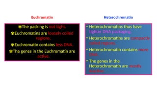
  • 28. Euchromatin ✾The packing is not tight. ✾Euchromatins are loosely coiled regions. ✾Euchromatin contains less DNA. ✾The genes in the Euchromatin are active. Heterochromatin • Heterochromatins thus have tighter DNA packaging. • Heterochromatins are compactly coiled regions. • Heterochromatin contains more DNA. • The genes in the Heterochromatin are mostly inactive. © Mr. Suman Neerukonda
  • 29. Mitosis The cell cycle is the regular sequence of events that takes place between one cell division and the next. It has three phases namely interphase, nuclear division and cell division.
  • 30. ✢ Interphase is often included in discussions of mitosis, but interphase is technically not part of mitosis, but rather encompasses stages G1, S, and G2 of the cell cycle. ✢The stage of the cell cycle when a cell is preparing itself to duplicate is called interphase. ✢During interphase, the cell obtains nutrients, and duplicates its chromosomes. ✢During interphase, the chromosomes are found arranged in the nucleus and appear as a network of long, thin threads, called chromatin.
  • 31. Summary of the cell cycle G1 phase cell grows DNA is transcribed Protein is synthesized S phase DNA is replicated G2 phase Cell prepares for division Mitosis Cell nucleus divides Cytokinesis Cytoplasm divides
  • 32. S phase (S stands for synthesis of DNA) ❃In S phase, the cell synthesizes a complete copy of the DNA in its nucleus. ❃It also duplicates a microtubule-organizing structure called the centrosome. ❃The centrosomes help separate DNA during M phase.
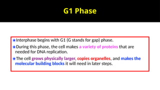
  • 33. G1 Phase ✬Interphase begins with G1 (G stands for gap) phase. ✬During this phase, the cell makes a variety of proteins that are needed for DNA replication. ✬The cell grows physically larger, copies organelles, and makes the molecular building blocks it will need in later steps.
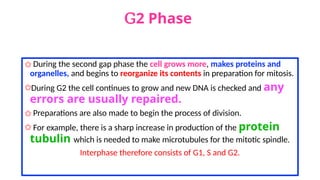
  • 34. G2 Phase ✩ During the second gap phase the cell grows more, makes proteins and organelles, and begins to reorganize its contents in preparation for mitosis. ✩During G2 the cell continues to grow and new DNA is checked and any errors are usually repaired. ✩ Preparations are also made to begin the process of division. ✩ For example, there is a sharp increase in production of the protein tubulin which is needed to make microtubules for the mitotic spindle. Interphase therefore consists of G1, S and G2.
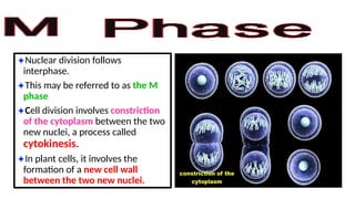
  • 36. ✦Nuclear division follows interphase. ✦This may be referred to as the M phase ✦Cell division involves constriction of the cytoplasm between the two new nuclei, a process called cytokinesis. ✦In plant cells, it involves the formation of a new cell wall between the two new nuclei.
  • 37. Mitosis is divided into four phases. Each phase is characterized by specific processes involving different structures. The four stages are called: Prophase Metaphase Anaphase Telophase
  • 38. Early Prophase centrosomes ✬They are only found in eukaryotic cells. ✬Centrosomes are comprised of two centrioles that are essentially just rings of microtubules. ✬The purpose of the centrosome is to help organize microtubules to be utilized during cell division.
  • 44. Mitosis and cytokinesis produce two genetically identical daughter cells.
  • 45. Centromere ❋a centromere is responsible for the movement of the replicated chromosomes into the two daughter cells during mitosis and meiosis. ❋Centromere joins the sister chromatids. ❋ The two copies of a replicated chromosome are called sister chromatids, and they must stay joined together until it is time for them to be physically pulled into the two future daughter cells.
  • 46. kinetochores are platelike structures composed of several layers. Multiple microtubules appear to insert into the kinetochore, which is positioned on the side of the chromosome facing the spindle pole.
  • 47. Centrosome ✺Centrosomes are comprised of two centrioles that are essentially just rings of microtubules. ✺The purpose of the centrosome is to help organize microtubules to be utilized during cell division. ✺The centrosome is a microtubule organizing center and consists of two centrioles that are held perpendicular to one another.
  • 48. ✽Most plants do not contain centrioles, but instead have microtubule clusters that function to direct the distribution of chromosomes. ✽ They also participate in splitting the cell during cytokinesis. ✽ During prophase, the plant cell begins to produce spindles from the organizing centers that grow into the nuclear region and attach to the chromosomes. ✽From there, they orchestrate the organization and segregation of chromosomes between daughter cells during mitosis. ✽The nuclear envelope itself appears to function as the main MTOC for microtubule nucleation and spindle organization during plant cell mitosis.
  • 49. Biological Significance of Mitosis ✼ It is an equational division through which identical daughter cells are produced having the same amount and type of genetic constitution as that of the parent cell. ✼It is responsible for growth and development of multi-cellular organisms from a single-celled zygote. ✼The number of chromosomes remains the same in all the cells produced by this division. ✼Thus, the daughter cells retain the same characters as those of the parent cell.
  • 50. ✼Mitosis helps in restoring wear and tear in body tissues, replacement of damaged or lost part, healing of wounds and regeneration of detached parts (as in tail of a lizards). ✼Immune response - The cloning of B- and T-lymphocytes during the immune response is dependent on mitosis. © Mr. Suman Neerukonda
  • 51. Asexual reproduction- It is a method of multiplication in unicellular organisms.
  • 52. The Significance of Telomeres
  • 53. ✺Telomere (tel-uh-meer) from the Greek telos (end) and meros (part). ✺The ends of chromosomes are ‘sealed’ by structures called telomeres. ✺These are made of DNA with short base sequences that are repeated many times. ✺In telomeres, one strand of the DNA is rich in the base guanine (G) and the other strand is rich in the complementary base cytosine (C). © Mr. Suman Neerukonda
  • 54. ✾Their main function is to ensure that when DNA is replicated, the ends of the molecule are included in the replication and not left out. ✾It has been shown that some cells do not ‘top up’ their telomeres at each division. ✾These tend to be fully differentiated cells. ✾With each division, their telomeres get a little shorter until the vital DNA is no longer protected and the cell dies. ✾This could be one of the mechanisms of ageing, by which we grow old and die.
  • 55. Stem cells ✫Stem cells have the remarkable potential to develop into many different cell types in the body during early life and growth. ✫A stem cell is a cell that can divide an unlimited number of times (by mitosis). ✫When it divides, each new cell has the potential to remain a stem cell or to develop (differentiate) into a specialized cell such as a blood cell or muscle cell.
  • 56. ✶Cell potency is a cell's ability to differentiate into other cell types. ✶The more cell types a cell can differentiate into, the greater its potency. ✶Totipotency is the ability of a single cell to divide and produce all of the differentiated cells in an organism. ✶Spores and zygotes are examples of totipotent cells
  • 57. One-celled fertilized egg ❂This cell is totipotent, meaning it has the potential to give rise to any and all human cells, such as brain, liver, blood or heart cells. ❂It can even give rise to an entire functional organism. ❂The first few cell divisions in embryonic development produce more totipotent cells. ❂After four days of embryonic cell division, the cells begin to specialize into pluripotentstem cells.
  • 58. Pluripotent Stem Cells ✫Pluripotent stem cells are master cells. ✫These cells have the potential of taking on many fates in the body, including all of the more than 200 different cell types. ✫Embryonic stem cells come from pluripotent cells, which exist only at the earliest stages of embryonic development. ✫ In humans, these cells no longer exist after about five days of development.
  • 59. ✬However, for growth and repair it is essential that small populations of stem cells remain which can produce new cells. ✬Adult stem cells have already lost some of the potency associated with embryonic stem cells and are no longer pluripotent. ✬They are only able to produce a few types of cell and may be described as multi potent. ✬For example, the stem cells found in bone marrow are of this type. ✬They can replicate any number of times, but can produce only blood cells, such as red blood cells, monocytes, neutrophils and lymphocytes. ✬Mature blood cells have a relatively short life span, so the existence of these stem cells is essential.
  • 60. ✾On the fourth day of development, the embryo forms into two layers, an outer layer which will become the placenta, and an inner mass which will form the tissues of the developing human body. ✾These inner cells, though they can form nearly any human tissue, cannot do so without the outer layer; so are not totipotent, but pluripotent. ✾As these pluripotent stem cells continue to divide, they begin to specialize further. © Mr. Suman Neerukonda
  • 61. Multipotent Stem Cells ✧These are less plastic and more differentiated stem cells. ✧They give rise to a limited range of cells within a tissue type. ✧The offspring of the pluripotent cells become the progenitors of such cell lines as blood cells, skin cells and nerve cells. ✧At this stage, they are multi potent. ✧They can become one of several types of cells within a given organ. ✧For example, multi potent blood stem cells can develop into red blood cells, white blood cells or platelets.
  • 62. ★An adult stem cell is a multi potent stem cell in adult humans that is used to replace cells that have died or lost function. ★ It is an undifferentiated cell present in differentiated tissue. ★ It renews itself and can specialize to yield all cell types present in the tissue from which it originated. ★So far, adult stem cells have been identified for many different tissue types such as hematopoietic (blood), neural, endothelial, muscle, mesenchymal, gastrointestinal, and epidermal cells.
  • 64. Stem Cell Therapy ❁Stem-cell therapy is the use of stem cells to treat or prevent a disease or condition. ❁Bone marrow transplantation is the only form of this therapy that has progressed beyond the experimental stage into routine medical practice, but in the future it is hoped to be able to treat conditions like diabetes, muscle and nerve damage, and brain disorders such as Parkinson’s and Huntington’s diseases. ❁Experiments with growing new tissues, or even organs, from isolated stem cells in the laboratory have also been conducted.
  • 65. ✭A disease in which abnormal cells divide uncontrollably and destroy body tissue. ✭Cancer develops when the body’s normal control mechanism stops working. ✭Old cells do not die and cells grow out of control, forming new, abnormal cells. ✭ These extra cells may form a mass of tissue, called a Tumor. © Mr. Suman Neerukonda
  • 68. ✩Cancers show us the importance of controlling cell division precisely, because cancers are a result of uncontrolled mitosis. ✩Cancerous cells divide repeatedly and form a tumor, which is an irregular mass of cells.
  • 69. ✺Cancers are thought to start when changes occur in the genes that control cell division. ✺A change in any gene is called a Mutation. ✺The particular term for a mutated gene that causes cancer is an oncogene, after the Greek word ‘onkos’, meaning bulk or mass.
  • 70. ✭Mutations are not unusual events, and most of the time they don’t lead to cancer. ✭Most mutated cells are either affected in some way that results in their early death or are destroyed by the body’s immune system. ✭Since most cells can be replaced, mutation usually has no harmful effect on the body. © Mr. Suman Neerukonda
  • 71. ✫Cancerous cells, however, manage to escape both possible fates, so, although the mutation may originally occur in only one cell, it is passed on to all that cell’s descendants. ✫By the time it is detected, a typical tumor usually contains about a thousand million cancerous cells. ✫Any agent that causes cancer is called a carcinogen and is described as carcinogenic.
  • 72. Benign Tumors ✹ A benign tumor does not invade nearby tissue or spread to other parts of the body the way cancer can. ✹ But benign tumors can be serious if they press on vital structures such as blood vessels or nerves. ✹ Therefore, sometimes they require treatment and other times they do not.
  • 73. Malignant Tumors ✬They spread through the body, invade other tissues, destroy them and cause cancer, and these are known as Malignant tumors. ✬Malignant tumors interfere with the normal functioning of the area where they have started to grow. ✬They may block the intestines, lungs or blood vessels. ✬Cells can break off and spread through the blood and lymphatic system to other parts of the body to form secondary growths. © Mr. Suman Neerukonda
  • 75. Metastasis ✭Metastasis is the spread of a cancer from one organ or part of the body to another without being directly connected with it. ✭In metastasis, cancer cells break away from the original (primary) tumor, travel through the blood or lymph system, and form a new tumor in other organs or tissues of the body. ✭The new, metastatic tumor is the same type of cancer as the primary tumor. ✭For example, if breast cancer spreads to the lung, the cancer cells in the lung are breast cancer cells, not lung cancer cells. ✭ The plural form of metastasis is metastases.
  • 77. © Mr. Suman Neerukonda