SlideShare a Scribd company logo
KONGUNADU COLLEGE OF ENGINEERING AND TECHNOLOGY
(AUTONOMOUS)
NAMAKKAL- TRICHY MAIN ROAD, THOTTIAM
DEPARTMENT OF BIOMEDICAL ENGINEERING
20BM708PE – NEURAL ENGINEERING
(REGULATION-KNCET - UGR2020)
UNIT III – EVOKED POTENTIALS
Ms. M. Thendral,
Assistant Professor / BME
KNCET
UNIT III - EVOKED POTENTIALS
•Evoked Potentials and Related Techniques: Visual
Evoked potentials (VEPS), Electroretinography and
other diagnostic approaches to the Visual System,
VEPS in infants and children, Somatosensory evoked
potentials, Diagnostic and Therapeutic role of
Magnetic stimulation in neurology
EVOKED POTENTIALS AND RELATED TECHNIQUES:
Evoked potential
• Evoked potential tests measure the electrical activity in areas of brain and spinal cord in response to certain stimuli. The
tests involve electrodes placed on specific parts of your scalp and/or other parts of your body and delivery of a stimulus (such as
images, sounds or electrical pulses). The electrodes capture brain’s and nerves’ electrical signal responses to the stimulus. Evoked
potential tests record how quickly and completely nerve signals reach the brain. They can find damage along nerve and brain
pathways.
Working of evoked potential
• An evoked potential test involves electrodes placed on specific parts of scalp and delivery of a stimulus. It can record the
brain’s electrical response to the stimulus. Nervous system consists of a vast network of nerves that send electrical signals to and
from other cells, glands and muscles all over the body. These nerves receive information from brain. The brain interprets the
information and controls the response.
• In an evoked potential test, electrodes on your scalp measure electrical signals (impulses) as they travel between brain cells. As
you experience the stimulus — either a visual pattern, series of sounds or very mild electric pulses or “shocks” —the nervous
system responds to and interprets the stimulus. The electrodes on scalp record the electrical activity of brain that results. The
evoked potential machine averages the EEG signals following multiple stimuli so that it can assess the functioning of a specific
neurological system. The evoked potential machine records the electrical response to the stimulation on several channels or traces.
The brain has specific waveforms that occur at very specific times in response to a various stimulus
EVOKED POTENTIALS
BLOCK DIAGRAM OF EVOKED
POTENTIALS
TYPES OF EVOKED POTENTIALS
1. Motor evoked potentials: Motor evoked potentials (MEPs)
are the electrical signals recorded from the descending motor
pathways or from muscles following stimulation of motor
pathways within the brain.
2. Sensory evoked potentials : Sensory evoked potentials
studies measure electrical activity in the brain in response to
stimulation of sight, sound, or touch. When the brain is
stimulated by sight, sound, or touch, signals travel along the
nerves to the brain. There, electrodes detect the signals and
display them. Sensory evoked potentials studies involve 3 tests
that measure response to visual, auditory, and electrical stimuli.
a. Visual evoked potential (VEP): A visual evoked potential
(VEP) test measures the electrical signal your visual cortex (a
region of brain) generates in response to visual stimulation. The
test is also called a visual evoked response (VER).
b. Brainstem auditory evoked response (BAER): Brainstem
auditory evoked potentials (BAEPs), also called brainstem auditory
evoked responses (BAERs), are very small
auditory evoked potentials in response to an auditory stimulus,
which are recorded by electrodes placed on the scalp.
c. Somatosensory evoked potentials (SEP): Somatosensory
evoked potential (SEP or SSEP) is the electrical activity of the
brain that results from the stimulation of touch. SEP tests are non-
invasive to assess the somatosensory system functioning.
3. Event related potentials: Event-related potentials (ERPs) are
electrical brain responses that occur in response to specific sensory,
cognitive, or motor events. They are measured using
electroencephalography (EEG) and are characterized by their time-
locked nature, meaning they occur at precise moments in response
to a particular stimulus or event.
VISUAL EVOKED POTENTIAL
• A visual evoked potential (VEP) test measures the electrical
signal your visual cortex (a region of brain) generates in
response to visual stimulation. The test is also called a visual
evoked response (VER).
• More specifically, a VEP test assesses the function of your
visual pathway, which includes your: Eyes, Optic nerves,
Optic tract, Optic radiation (the part of your visual pathway
that transmits visual input coming from your retina, optic
nerve and optic tract) and Cerebral cortex
TYPES OF VEP
Flash VEP
• Flash Visual Evoked
Potential (VEP) is a test
used to assess the function
of the visual pathways in
the brain.
• It involves presenting a
series of flashing light
stimuli to a person's eyes
and recording the electrical
responses generated by the
visual system.
PATTERN REVERSAL VEP
• It is a diagnostic test that
measures the electrical
activity in the brain in
response to visual stimuli.
• Specifically, it focuses on
the brain's response to
alternating patterns of
black and white squares or
lines that rapidly switch
back and forth (reversal).
PATTERN ONSET OFFSET VEP
• It focuses on the brain's
response to the onset and
offset of repeating visual
patterns.
• This test involves
presenting patterns, such as
checkerboards to a person's
eyes in a repeating
sequence while recording
the electrical responses
generated by the visual
system.
Block diagram of VEP
• The patient is seated at an appropriate distance from the visual
stimulus.
• The appropriate recording (active) electrode is attached to the
posterior scalp and connected to the positive input of the
differential amplifier.
• A similar reference electrode is attached to a visually neutral
site on the head (e.g., the earlobe) and connected to the
negative input of the differential amplifier.
• A ground (GND) electrode is attached to the forehead. A
stimulus generator is used to select the desired stimulus type
(flash, pattern reversal, or pattern onset-offset), temporal
frequency, pattern type and size of the pattern elements.
• The stimulus generator also sends a signal to the signal
averaging computer that is synchronous with the stimulus
presentation and triggers each averaging epoch.
• The signal averaging computer usually controls the duration of
the averaging epoch, the number of averages collected, the
signal conditioning, and signal analysis.
ELECTRORETINOGRAM
• An electroretinogram (ERG) is a diagnostic test used to measure
the electrical activity of the retina in response to light stimulation.
• It is a valuable tool for assessing the function of the retina and
identifying any abnormalities or disorders affecting the visual
system.
• The normal response pattern has waves called a wave and b wave.
• A-wave: Both rods and cones contributes to it. This is produce
due to the hyper-polarisation of rod and cones
• B-wave: The b-wave is generated by ON and OFF bipolar cell (B)
activity.
ERG
• Light Stimulator: During an ERG test, the patient is exposed to flashes of
light or patterns of light that are carefully controlled in intensity and duration.
• Electrodes: Electrodes are placed on the surface of the eye or around the eye
to detect the electrical responses of the retina. The most commonly used
electrodes are corneal electrodes (contact lens-type electrodes) or skin
electrodes placed on the skin around the eye.
• Retinal Response: When the retina is exposed to light, the photoreceptor
cells (rods and cones) in the retina are stimulated and produce electrical
responses. These responses are transmitted through the optic nerve to the
visual processing centres in the brain.
• Amplification: The electrical responses from the electrodes are extremely
small and require amplification for accurate measurement.
• Filtering: The amplified electrical response may contain noise or artifacts that
need to be filtered.
• Analog-to-Digital Converter (ADC): The analog signals are then converted
into digital signals using an ADC.
• Data Display: The recorded data is analysed to assess the overall health and
function of the retina.
OTHER DIAGNOSTIC APPROACHES TO THE
VISUAL SYSTEM
ELECTROOCULOGRAPHY
 Electrooculography (EOG) is a non-invasive technique used to measure the electrical activity generated by the muscles surrounding
the eyes.
 The electrical signals produced by eye movements are recorded through electrodes placed on the skin near the eyes.
 EOG is widely used in various applications, such as medical diagnostics, sleep studies, eye movement studies, and brain-computer
interface research.
PRINCIPLE OF EOG:
 EOG works based on the principle that the eyes have a positively charged cornea and a negatively charged retina, creating a dipole
between them.
 When the eyes move, the distribution of these charges changes and this generates electrical potentials around the eyes.
 By measuring these changes in electrical potentials using electrodes, we can determine the direction and magnitude of eye
movements.
RECORDING OF EOG
VEPS IN INFANTS AND CHILDREN
•Visual evoked potentials (VEPs) are a type of electrophysiological measurement used to assess the
visual system's response to visual stimuli. In infants, VEPs can provide insights into the development of their
visual pathways and help identify potential issues with visual processing.
•The process involves presenting visual stimuli, like patterns or flashing lights, while recording the
brain's electrical activity through electrodes placed on the scalp. VEPs can aid in diagnosing visual
impairments, tracking visual maturation, and evaluating the effectiveness of treatments.
• Stimulus Presentation: Visual stimuli, such as patterns or flashing lights, are presented to the subject. These
stimuli are designed to evoke a response from the visual system
• Electrode Placement: Electrodes are attached to the scalp in specific locations to record the brain's electrical
activity. Common electrode placements include those over the visual cortex at the back of the head.
• Amplification: The weak electrical signals picked up by the electrodes are amplified to make them
measurable and more easily distinguishable from noise.
VEPS IN INFANTS AND CHILDREN
• Signal Processing: The amplified signals undergo signal
processing, including filtering and noise reduction, to enhance the
quality of the recorded data.
• Averaging: Multiple repetitions of the same stimulus are presented
and the recorded brain responses are averaged to improve the signal-
to-noise ratio, as the brain's response is often a small signal
compared to background noise.
• Data Analysis: The averaged and processed signals are analysed to
identify specific components of the brain's response related to the
visual stimuli. This involves identifying peaks and troughs in the
recorded waveform.
• Results Interpretation: The obtained VEP waveform is interpreted
by comparing it to known patterns and characteristics associated
with normal or abnormal visual processing. Expertise is required to
accurately interpret the results, especially in the case of infants.
SOMATOSENSORY EVOKED POTENTIALS
Somatosensory evoked potentials (SSEPs) are electrical signals generated by the nervous system in response to
sensory stimuli applied to the body.
1. Median nerve sensory evoked potentials
• Median nerve sensory evoked potentials (SEP) are
a diagnostic test used to assess the function of the
median nerve, typically at the wrist. This test
involves placing electrodes on the skin to record
electrical signals generated by the sensory nerve
fibers in response to a controlled stimulus applied
to the median nerve. It can help in diagnosing
conditions such as carpal tunnel syndrome, which
can cause impaired sensory function in the hand
and wrist. The results of this test provide valuable
information about the integrity and function of the
median nerve.
SOMATOSENSORY EVOKED POTENTIALS
• Tibial nerve sensory evoked potentials
• Tibial nerve sensory evoked potentials (SEPs) are
diagnostic tests used to assess the function of the
tibial nerve, typically at the ankle or lower leg.
Similar to other SEP tests, electrodes are placed on
the skin to record electrical signals generated by the
sensory nerve fibres in response to a controlled
stimulus applied to the tibial nerve. Tibial nerve
SEPs can help diagnose conditions related to nerve
damage or compression affecting the tibial nerve,
such as peripheral neuropathy or radiculopathy.
These test results provide insights into the integrity
and function of the tibial nerve.
SOMATOSENSORY
EVOKED POTENTIALS
• Stimulus Delivery: A controlled sensory stimulus such as an
electrical stimulus is applied to the body, typically to the limbs.
• Electrode Placement: Electrodes are placed on the scalp to record
the electrical signals generated by the nervous system in response
to the stimulus.
• Amplification and Filtering: The recorded signals are amplified
and filtered to enhance the quality of the signals and remove
unwanted noise.
• Signal Averaging: Multiple trials of stimulus presentation are
averaged together to enhance the signal-to-noise ratio.
• Data Analysis: The averaged responses are analysed to identify the
various components of the SSEP waveform, such as peaks and
latencies.
• Interpretation: The SSEP findings are interpreted by comparing
them to established norms or previous recordings, aiding in
diagnosis or assessment.
DIAGNOSTIC AND THERAPEUTIC ROLE OF MAGNETIC
STIMULATION IN NEUROLOGY
• Magnetic stimulation, like transcranial magnetic stimulation (TMS), plays a significant diagnostic and
therapeutic role in neurology. It's used to non-invasively stimulate or modulate brain activity. In diagnostics,
TMS can map brain function and connectivity, aiding in conditions like epilepsy and stroke. Therapeutically,
it's used for depression, migraines, and certain movement disorders
• Transcranial Magnetic Stimulation (TMS) is a non-invasive neurostimulation technique that uses
electromagnetic induction to generate brief, focused magnetic pulses, which can penetrate the skull and
induce electrical currents in specific regions of the brain. TMS has both diagnostic and therapeutic
applications in the field of neurology.
Diagnostic Procedure:
• Patient Assessment: Evaluate the patient's medical history, neurological symptoms, and condition to
determine the need for diagnostic magnetic stimulation.
• Preparation: Position the patient comfortably in a controlled environment, ensuring proper safety measures
are in place.
• Brain Mapping: Use Transcranial Magnetic Stimulation (TMS) to stimulate different areas of the brain's
cortex. Measure motor responses or evoked potentials in specific muscles to map brain function and
connectivity.
• Motor Threshold Determination: Identify the motor threshold, the intensity required to evoke a motor
response. This helps calibrate subsequent stimulations.
• Functional Imaging: Combine TMS with techniques like functional MRI (fMRI) to understand brain
connectivity and networks in conditions like epilepsy or stroke.
Therapeutic Procedure:
Patient Evaluation: Assess the patient's suitability for therapeutic magnetic stimulation based on their medical history, current medications,
and condition.
Preparation: Place the patient in a comfortable position, ensuring their safety during the procedure.
Target Selection: Identify the specific brain region to be targeted based on the therapeutic goal (e.g., depression, migraine relief, movement
disorder management).
Stimulation Parameters: Determine the appropriate stimulation intensity, frequency, and duration for the specific condition and target area.
Repetitive Transcranial Magnetic Stimulation (rTMS):
• For depression or migraine treatment, position the coil over the prefrontal cortex or other relevant regions.
• Administer a series of magnetic pulses at the specified frequency and intensity over several sessions.
Single-Pulse TMS or Deep Brain Stimulation (DBS) (for Movement Disorders):
Administer single pulses or repetitive pulses targeting motor cortex or deep brain structures involved in movement control.
Monitoring and Adjustment:
• Monitor the patient's responses and potential side effects during and after each session.
• Adjust stimulation parameters based on patient feedback and progress.

More Related Content

PPT
Biomedical Signals
PPTX
NEUROPHYSIOLOGY technique.pptx
PPTX
INTRODUCTION TO BIOMEDICAL SIGNALS in BSP
PPT
SUMSEM-2021-22_ECE6007_ETH_VL2021220701295_Reference_Material_I_04-07-2022_EE...
PPTX
Presentation EEG (1).pptx
PPTX
Eeg seminar
PDF
Clinical teaching on electroencephelography
PDF
Eeg presentation for studying and understanding
Biomedical Signals
NEUROPHYSIOLOGY technique.pptx
INTRODUCTION TO BIOMEDICAL SIGNALS in BSP
SUMSEM-2021-22_ECE6007_ETH_VL2021220701295_Reference_Material_I_04-07-2022_EE...
Presentation EEG (1).pptx
Eeg seminar
Clinical teaching on electroencephelography
Eeg presentation for studying and understanding

Similar to NEURAL ENGINEERING UNIT 3 EVOKED POTENTIAL (20)

PDF
International Journal of Computational Engineering Research(IJCER)
PDF
International Journal of Computational Engineering Research(IJCER)
PPTX
Basics of EEG
PPTX
Electroencephal ogram (eeg)
PPTX
Application of eeg wave
PPTX
Brain Control Interface using ELECTROENCEPHALOGRAPHY
PDF
A0510107
PDF
Biomarker and human cognition.pdf
PPTX
NEURAL ENGINEERING UNIT II ELECTROENCEPHALOGRAPHY
PDF
Textbook of electroencephalography
PPTX
MEDICAL ELECTRONICS_EC8073_Unit 1session 8
PDF
Feature Extraction Techniques and Classification Algorithms for EEG Signals t...
PPTX
Recording brain activity
PPTX
Evoked Potentials.pptx
PPTX
Eeg presentation
PDF
PPTX
lecture2 EEG EMG VCG PCG_1105315566.pptx
PPTX
EEG & EMG new.pptx
International Journal of Computational Engineering Research(IJCER)
Basics of EEG
Electroencephal ogram (eeg)
Application of eeg wave
Brain Control Interface using ELECTROENCEPHALOGRAPHY
A0510107
Biomarker and human cognition.pdf
NEURAL ENGINEERING UNIT II ELECTROENCEPHALOGRAPHY
Textbook of electroencephalography
MEDICAL ELECTRONICS_EC8073_Unit 1session 8
Feature Extraction Techniques and Classification Algorithms for EEG Signals t...
Recording brain activity
Evoked Potentials.pptx
Eeg presentation
lecture2 EEG EMG VCG PCG_1105315566.pptx
EEG & EMG new.pptx
Ad

More from thendralm1 (6)

PPTX
BIOMEDICAL IMSTRUMENTION UNIT III SIGNAL CONDITIONING CIRCUITS
PPTX
ELECTROPHYSIOLOGICAL EVALUATION IN SPECIAL SITUATIONS
PPTX
NEURAL ENGINEERING UNIT 4 FUNCTIONAL NEUROIMAGING AND RECOGNITION
PPTX
UNIT I NERVE EXCITABILITY AND ELECTROMYOGRAPHY
PPTX
BIOMEDICAL INSTRUMENTATION UNIT 2 BIOPOTENTIAL MEASUREMENTS
PPTX
BIOMEDICAL INSTRUMENTATION UNIT I BIOPOTENTIAL ELECTRODES
BIOMEDICAL IMSTRUMENTION UNIT III SIGNAL CONDITIONING CIRCUITS
ELECTROPHYSIOLOGICAL EVALUATION IN SPECIAL SITUATIONS
NEURAL ENGINEERING UNIT 4 FUNCTIONAL NEUROIMAGING AND RECOGNITION
UNIT I NERVE EXCITABILITY AND ELECTROMYOGRAPHY
BIOMEDICAL INSTRUMENTATION UNIT 2 BIOPOTENTIAL MEASUREMENTS
BIOMEDICAL INSTRUMENTATION UNIT I BIOPOTENTIAL ELECTRODES
Ad

Recently uploaded (20)

PPTX
Fundamentals of Mechanical Engineering.pptx
PDF
Human-AI Collaboration: Balancing Agentic AI and Autonomy in Hybrid Systems
PPT
Introduction, IoT Design Methodology, Case Study on IoT System for Weather Mo...
PPTX
Information Storage and Retrieval Techniques Unit III
PDF
Analyzing Impact of Pakistan Economic Corridor on Import and Export in Pakist...
PDF
keyrequirementskkkkkkkkkkkkkkkkkkkkkkkkkkkkkkkkkkkkk
PPTX
Nature of X-rays, X- Ray Equipment, Fluoroscopy
PDF
COURSE DESCRIPTOR OF SURVEYING R24 SYLLABUS
PDF
BIO-INSPIRED ARCHITECTURE FOR PARSIMONIOUS CONVERSATIONAL INTELLIGENCE : THE ...
PPTX
CURRICULAM DESIGN engineering FOR CSE 2025.pptx
PDF
Unit I ESSENTIAL OF DIGITAL MARKETING.pdf
PDF
Automation-in-Manufacturing-Chapter-Introduction.pdf
PPTX
Fundamentals of safety and accident prevention -final (1).pptx
PDF
UNIT no 1 INTRODUCTION TO DBMS NOTES.pdf
PDF
Integrating Fractal Dimension and Time Series Analysis for Optimized Hyperspe...
PDF
The CXO Playbook 2025 – Future-Ready Strategies for C-Suite Leaders Cerebrai...
PPTX
UNIT 4 Total Quality Management .pptx
PDF
Categorization of Factors Affecting Classification Algorithms Selection
PPTX
Current and future trends in Computer Vision.pptx
PPT
A5_DistSysCh1.ppt_INTRODUCTION TO DISTRIBUTED SYSTEMS
Fundamentals of Mechanical Engineering.pptx
Human-AI Collaboration: Balancing Agentic AI and Autonomy in Hybrid Systems
Introduction, IoT Design Methodology, Case Study on IoT System for Weather Mo...
Information Storage and Retrieval Techniques Unit III
Analyzing Impact of Pakistan Economic Corridor on Import and Export in Pakist...
keyrequirementskkkkkkkkkkkkkkkkkkkkkkkkkkkkkkkkkkkkk
Nature of X-rays, X- Ray Equipment, Fluoroscopy
COURSE DESCRIPTOR OF SURVEYING R24 SYLLABUS
BIO-INSPIRED ARCHITECTURE FOR PARSIMONIOUS CONVERSATIONAL INTELLIGENCE : THE ...
CURRICULAM DESIGN engineering FOR CSE 2025.pptx
Unit I ESSENTIAL OF DIGITAL MARKETING.pdf
Automation-in-Manufacturing-Chapter-Introduction.pdf
Fundamentals of safety and accident prevention -final (1).pptx
UNIT no 1 INTRODUCTION TO DBMS NOTES.pdf
Integrating Fractal Dimension and Time Series Analysis for Optimized Hyperspe...
The CXO Playbook 2025 – Future-Ready Strategies for C-Suite Leaders Cerebrai...
UNIT 4 Total Quality Management .pptx
Categorization of Factors Affecting Classification Algorithms Selection
Current and future trends in Computer Vision.pptx
A5_DistSysCh1.ppt_INTRODUCTION TO DISTRIBUTED SYSTEMS

NEURAL ENGINEERING UNIT 3 EVOKED POTENTIAL

  • 1. KONGUNADU COLLEGE OF ENGINEERING AND TECHNOLOGY (AUTONOMOUS) NAMAKKAL- TRICHY MAIN ROAD, THOTTIAM DEPARTMENT OF BIOMEDICAL ENGINEERING 20BM708PE – NEURAL ENGINEERING (REGULATION-KNCET - UGR2020) UNIT III – EVOKED POTENTIALS Ms. M. Thendral, Assistant Professor / BME KNCET
  • 2. UNIT III - EVOKED POTENTIALS •Evoked Potentials and Related Techniques: Visual Evoked potentials (VEPS), Electroretinography and other diagnostic approaches to the Visual System, VEPS in infants and children, Somatosensory evoked potentials, Diagnostic and Therapeutic role of Magnetic stimulation in neurology
  • 3. EVOKED POTENTIALS AND RELATED TECHNIQUES: Evoked potential • Evoked potential tests measure the electrical activity in areas of brain and spinal cord in response to certain stimuli. The tests involve electrodes placed on specific parts of your scalp and/or other parts of your body and delivery of a stimulus (such as images, sounds or electrical pulses). The electrodes capture brain’s and nerves’ electrical signal responses to the stimulus. Evoked potential tests record how quickly and completely nerve signals reach the brain. They can find damage along nerve and brain pathways. Working of evoked potential • An evoked potential test involves electrodes placed on specific parts of scalp and delivery of a stimulus. It can record the brain’s electrical response to the stimulus. Nervous system consists of a vast network of nerves that send electrical signals to and from other cells, glands and muscles all over the body. These nerves receive information from brain. The brain interprets the information and controls the response. • In an evoked potential test, electrodes on your scalp measure electrical signals (impulses) as they travel between brain cells. As you experience the stimulus — either a visual pattern, series of sounds or very mild electric pulses or “shocks” —the nervous system responds to and interprets the stimulus. The electrodes on scalp record the electrical activity of brain that results. The evoked potential machine averages the EEG signals following multiple stimuli so that it can assess the functioning of a specific neurological system. The evoked potential machine records the electrical response to the stimulation on several channels or traces. The brain has specific waveforms that occur at very specific times in response to a various stimulus
  • 4. EVOKED POTENTIALS BLOCK DIAGRAM OF EVOKED POTENTIALS TYPES OF EVOKED POTENTIALS
  • 5. TYPES OF EVOKED POTENTIALS 1. Motor evoked potentials: Motor evoked potentials (MEPs) are the electrical signals recorded from the descending motor pathways or from muscles following stimulation of motor pathways within the brain. 2. Sensory evoked potentials : Sensory evoked potentials studies measure electrical activity in the brain in response to stimulation of sight, sound, or touch. When the brain is stimulated by sight, sound, or touch, signals travel along the nerves to the brain. There, electrodes detect the signals and display them. Sensory evoked potentials studies involve 3 tests that measure response to visual, auditory, and electrical stimuli. a. Visual evoked potential (VEP): A visual evoked potential (VEP) test measures the electrical signal your visual cortex (a region of brain) generates in response to visual stimulation. The test is also called a visual evoked response (VER). b. Brainstem auditory evoked response (BAER): Brainstem auditory evoked potentials (BAEPs), also called brainstem auditory evoked responses (BAERs), are very small auditory evoked potentials in response to an auditory stimulus, which are recorded by electrodes placed on the scalp. c. Somatosensory evoked potentials (SEP): Somatosensory evoked potential (SEP or SSEP) is the electrical activity of the brain that results from the stimulation of touch. SEP tests are non- invasive to assess the somatosensory system functioning. 3. Event related potentials: Event-related potentials (ERPs) are electrical brain responses that occur in response to specific sensory, cognitive, or motor events. They are measured using electroencephalography (EEG) and are characterized by their time- locked nature, meaning they occur at precise moments in response to a particular stimulus or event.
  • 6. VISUAL EVOKED POTENTIAL • A visual evoked potential (VEP) test measures the electrical signal your visual cortex (a region of brain) generates in response to visual stimulation. The test is also called a visual evoked response (VER). • More specifically, a VEP test assesses the function of your visual pathway, which includes your: Eyes, Optic nerves, Optic tract, Optic radiation (the part of your visual pathway that transmits visual input coming from your retina, optic nerve and optic tract) and Cerebral cortex
  • 8. Flash VEP • Flash Visual Evoked Potential (VEP) is a test used to assess the function of the visual pathways in the brain. • It involves presenting a series of flashing light stimuli to a person's eyes and recording the electrical responses generated by the visual system.
  • 9. PATTERN REVERSAL VEP • It is a diagnostic test that measures the electrical activity in the brain in response to visual stimuli. • Specifically, it focuses on the brain's response to alternating patterns of black and white squares or lines that rapidly switch back and forth (reversal).
  • 10. PATTERN ONSET OFFSET VEP • It focuses on the brain's response to the onset and offset of repeating visual patterns. • This test involves presenting patterns, such as checkerboards to a person's eyes in a repeating sequence while recording the electrical responses generated by the visual system.
  • 11. Block diagram of VEP • The patient is seated at an appropriate distance from the visual stimulus. • The appropriate recording (active) electrode is attached to the posterior scalp and connected to the positive input of the differential amplifier. • A similar reference electrode is attached to a visually neutral site on the head (e.g., the earlobe) and connected to the negative input of the differential amplifier. • A ground (GND) electrode is attached to the forehead. A stimulus generator is used to select the desired stimulus type (flash, pattern reversal, or pattern onset-offset), temporal frequency, pattern type and size of the pattern elements. • The stimulus generator also sends a signal to the signal averaging computer that is synchronous with the stimulus presentation and triggers each averaging epoch. • The signal averaging computer usually controls the duration of the averaging epoch, the number of averages collected, the signal conditioning, and signal analysis.
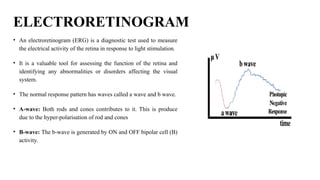
  • 12. ELECTRORETINOGRAM • An electroretinogram (ERG) is a diagnostic test used to measure the electrical activity of the retina in response to light stimulation. • It is a valuable tool for assessing the function of the retina and identifying any abnormalities or disorders affecting the visual system. • The normal response pattern has waves called a wave and b wave. • A-wave: Both rods and cones contributes to it. This is produce due to the hyper-polarisation of rod and cones • B-wave: The b-wave is generated by ON and OFF bipolar cell (B) activity.
  • 13. ERG • Light Stimulator: During an ERG test, the patient is exposed to flashes of light or patterns of light that are carefully controlled in intensity and duration. • Electrodes: Electrodes are placed on the surface of the eye or around the eye to detect the electrical responses of the retina. The most commonly used electrodes are corneal electrodes (contact lens-type electrodes) or skin electrodes placed on the skin around the eye. • Retinal Response: When the retina is exposed to light, the photoreceptor cells (rods and cones) in the retina are stimulated and produce electrical responses. These responses are transmitted through the optic nerve to the visual processing centres in the brain. • Amplification: The electrical responses from the electrodes are extremely small and require amplification for accurate measurement. • Filtering: The amplified electrical response may contain noise or artifacts that need to be filtered. • Analog-to-Digital Converter (ADC): The analog signals are then converted into digital signals using an ADC. • Data Display: The recorded data is analysed to assess the overall health and function of the retina.
  • 14. OTHER DIAGNOSTIC APPROACHES TO THE VISUAL SYSTEM ELECTROOCULOGRAPHY  Electrooculography (EOG) is a non-invasive technique used to measure the electrical activity generated by the muscles surrounding the eyes.  The electrical signals produced by eye movements are recorded through electrodes placed on the skin near the eyes.  EOG is widely used in various applications, such as medical diagnostics, sleep studies, eye movement studies, and brain-computer interface research. PRINCIPLE OF EOG:  EOG works based on the principle that the eyes have a positively charged cornea and a negatively charged retina, creating a dipole between them.  When the eyes move, the distribution of these charges changes and this generates electrical potentials around the eyes.  By measuring these changes in electrical potentials using electrodes, we can determine the direction and magnitude of eye movements.
  • 16. VEPS IN INFANTS AND CHILDREN •Visual evoked potentials (VEPs) are a type of electrophysiological measurement used to assess the visual system's response to visual stimuli. In infants, VEPs can provide insights into the development of their visual pathways and help identify potential issues with visual processing. •The process involves presenting visual stimuli, like patterns or flashing lights, while recording the brain's electrical activity through electrodes placed on the scalp. VEPs can aid in diagnosing visual impairments, tracking visual maturation, and evaluating the effectiveness of treatments. • Stimulus Presentation: Visual stimuli, such as patterns or flashing lights, are presented to the subject. These stimuli are designed to evoke a response from the visual system • Electrode Placement: Electrodes are attached to the scalp in specific locations to record the brain's electrical activity. Common electrode placements include those over the visual cortex at the back of the head. • Amplification: The weak electrical signals picked up by the electrodes are amplified to make them measurable and more easily distinguishable from noise.
  • 17. VEPS IN INFANTS AND CHILDREN • Signal Processing: The amplified signals undergo signal processing, including filtering and noise reduction, to enhance the quality of the recorded data. • Averaging: Multiple repetitions of the same stimulus are presented and the recorded brain responses are averaged to improve the signal- to-noise ratio, as the brain's response is often a small signal compared to background noise. • Data Analysis: The averaged and processed signals are analysed to identify specific components of the brain's response related to the visual stimuli. This involves identifying peaks and troughs in the recorded waveform. • Results Interpretation: The obtained VEP waveform is interpreted by comparing it to known patterns and characteristics associated with normal or abnormal visual processing. Expertise is required to accurately interpret the results, especially in the case of infants.
  • 18. SOMATOSENSORY EVOKED POTENTIALS Somatosensory evoked potentials (SSEPs) are electrical signals generated by the nervous system in response to sensory stimuli applied to the body. 1. Median nerve sensory evoked potentials • Median nerve sensory evoked potentials (SEP) are a diagnostic test used to assess the function of the median nerve, typically at the wrist. This test involves placing electrodes on the skin to record electrical signals generated by the sensory nerve fibers in response to a controlled stimulus applied to the median nerve. It can help in diagnosing conditions such as carpal tunnel syndrome, which can cause impaired sensory function in the hand and wrist. The results of this test provide valuable information about the integrity and function of the median nerve.
  • 19. SOMATOSENSORY EVOKED POTENTIALS • Tibial nerve sensory evoked potentials • Tibial nerve sensory evoked potentials (SEPs) are diagnostic tests used to assess the function of the tibial nerve, typically at the ankle or lower leg. Similar to other SEP tests, electrodes are placed on the skin to record electrical signals generated by the sensory nerve fibres in response to a controlled stimulus applied to the tibial nerve. Tibial nerve SEPs can help diagnose conditions related to nerve damage or compression affecting the tibial nerve, such as peripheral neuropathy or radiculopathy. These test results provide insights into the integrity and function of the tibial nerve.
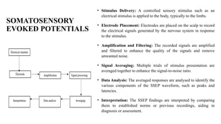
  • 20. SOMATOSENSORY EVOKED POTENTIALS • Stimulus Delivery: A controlled sensory stimulus such as an electrical stimulus is applied to the body, typically to the limbs. • Electrode Placement: Electrodes are placed on the scalp to record the electrical signals generated by the nervous system in response to the stimulus. • Amplification and Filtering: The recorded signals are amplified and filtered to enhance the quality of the signals and remove unwanted noise. • Signal Averaging: Multiple trials of stimulus presentation are averaged together to enhance the signal-to-noise ratio. • Data Analysis: The averaged responses are analysed to identify the various components of the SSEP waveform, such as peaks and latencies. • Interpretation: The SSEP findings are interpreted by comparing them to established norms or previous recordings, aiding in diagnosis or assessment.
  • 21. DIAGNOSTIC AND THERAPEUTIC ROLE OF MAGNETIC STIMULATION IN NEUROLOGY • Magnetic stimulation, like transcranial magnetic stimulation (TMS), plays a significant diagnostic and therapeutic role in neurology. It's used to non-invasively stimulate or modulate brain activity. In diagnostics, TMS can map brain function and connectivity, aiding in conditions like epilepsy and stroke. Therapeutically, it's used for depression, migraines, and certain movement disorders • Transcranial Magnetic Stimulation (TMS) is a non-invasive neurostimulation technique that uses electromagnetic induction to generate brief, focused magnetic pulses, which can penetrate the skull and induce electrical currents in specific regions of the brain. TMS has both diagnostic and therapeutic applications in the field of neurology.
  • 22. Diagnostic Procedure: • Patient Assessment: Evaluate the patient's medical history, neurological symptoms, and condition to determine the need for diagnostic magnetic stimulation. • Preparation: Position the patient comfortably in a controlled environment, ensuring proper safety measures are in place. • Brain Mapping: Use Transcranial Magnetic Stimulation (TMS) to stimulate different areas of the brain's cortex. Measure motor responses or evoked potentials in specific muscles to map brain function and connectivity. • Motor Threshold Determination: Identify the motor threshold, the intensity required to evoke a motor response. This helps calibrate subsequent stimulations. • Functional Imaging: Combine TMS with techniques like functional MRI (fMRI) to understand brain connectivity and networks in conditions like epilepsy or stroke.
  • 23. Therapeutic Procedure: Patient Evaluation: Assess the patient's suitability for therapeutic magnetic stimulation based on their medical history, current medications, and condition. Preparation: Place the patient in a comfortable position, ensuring their safety during the procedure. Target Selection: Identify the specific brain region to be targeted based on the therapeutic goal (e.g., depression, migraine relief, movement disorder management). Stimulation Parameters: Determine the appropriate stimulation intensity, frequency, and duration for the specific condition and target area. Repetitive Transcranial Magnetic Stimulation (rTMS): • For depression or migraine treatment, position the coil over the prefrontal cortex or other relevant regions. • Administer a series of magnetic pulses at the specified frequency and intensity over several sessions. Single-Pulse TMS or Deep Brain Stimulation (DBS) (for Movement Disorders): Administer single pulses or repetitive pulses targeting motor cortex or deep brain structures involved in movement control. Monitoring and Adjustment: • Monitor the patient's responses and potential side effects during and after each session. • Adjust stimulation parameters based on patient feedback and progress.