Cysts and Cyst-Like Lesions
Dr. Hadi Munib
Oral and Maxillofacial Surgery Resident
Introduction
▪ Cyst is a pathologic cavity filled with fluid, lined by epithelium, and surrounded by a
definite connective tissue wall.
▪ The cystic fluid either is secreted by the cells lining the cavity or derives from the
surrounding tissue fluid.
▪ Cysts occur more often in the jaws than in any other bone
▪ Cysts are radiolucent lesions, and the prevalent clinical features are swelling, lack of
pain (unless secondarily infected or is related to a non-vital tooth),
▪ Association with unerupted teeth, especially third molars.
Radiographic Features
▪ LOCATION
▪ Any location in the maxilla or mandible but are rare in the condyle and coronoid process.
▪ Odontogenic cysts are found most often in the tooth-bearing region.
▪ In mandible, they originate above the inferior alveolar nerve canal.
▪ Odontogenic cysts may grow into the maxillary antrum.
▪ PERIPHERY
▪ Well-Defined and corticated (fairly uniform, thin, radiopaque line).
▪ Secondary infection or a chronic state can change this appearance into a thicker, more sclerotic
boundary or make the cortex less apparent.
▪ SHAPE; Round or oval, resembling a fluid-filled balloon. Some cysts may have a scalloped
boundary.
Radiographic Features
▪ INTERNAL STRUCTURE; Totally radiolucent.
▪ However, long-standing cysts may have dystrophic calcification, which can give the internal
aspect a sparse, particulate appearance.
▪ Some cysts have septa, which produce multiple loculations separated by these bony walls or
septa.
▪ Cysts that have a scalloped periphery may appear to have internal septa
▪ EFFECTS ON SURROUNDING STRUCTURE
▪ Sometimes causing displacement and resorption of teeth. [Sharp and Curved]
▪ Cysts can expand the mandible, usually in a smooth, curved manner, and change the buccal or
lingual cortical plate into a thin cortical boundary.
▪ Cysts may displace the inferior alveolar nerve canal in an inferior direction or invaginate into
the maxillary antrum.
Cysts
Odontogenic
Non-Odontogenic
Radicular Cyst
▪ Periapical cyst, apical periodontal cyst, and dental cyst
▪ Epithelial cells (Malassez) in the periodontal ligament are stimulated to proliferate and
undergo cystic degeneration by inflammatory products from a non-vital tooth.
▪ Clinical Features
▪ Most common type of cyst in the jaws.
▪ No symptoms unless secondary infection occurs.
▪ May feel bony and hard if the cortex is intact, crepitant as the bone thins, and rubbery
and fluctuant if the outer cortex is lost.
▪ The incidence is greater in the third to sixth decades and shows a slight male
predominance.
Radicular Cyst
▪ Radiographic Features
▪ Location.
▪ At the apex of a non-vital tooth
▪ Occasionally it appears at the opening of an accessory canal, or infrequently in a deep
periodontal pocket.
▪ 60% are found in the maxilla, especially around incisors and canines.
▪ Well-defined cortical border and Radiolucent internal Structure
▪ Displacement/ Resorption of teeth
▪ Differential Diagnosis; Apical Granuloma
▪ Management
Radiographic Interpretation of Cyst and Cyst-like Lesions of the Jaws
Healing Cyst
Residual Cysts
▪ Cyst that remains after incomplete removal of the original cyst; radicular cyst that
may be left behind
▪ Clinical Features; Asymptomatic
▪ Radiographic Features
▪ Location. Found slightly more often in the mandible.
▪ In the mandible the epicenter is always above the inferior alveolar nerve canal
▪ A residual cyst has a cortical margin and is oval or circular. Radiolucent
▪ Effects on Surrounding Structures. can cause tooth displacement or resorption,
invagination into the maxillary antrum or depress the inferior alveolar nerve canal
Residual Cyst
▪ Differential Diagnosis
▪ Solitary Bone Cyst ; below the mandibular canal
▪ Odontogenic Keratocyst.
▪ Management
▪ Surgical removal or marsupialization
Dentigerous Cyst
▪ Follicular cyst
▪ Forms around the crown of an unerupted tooth.
▪ An eruption cyst is the soft tissue counterpart
▪ Dentigerous cysts are the second most common type of cyst
▪ Unerupted tooth or supernumerary tooth.
▪ Around supernumerary teeth account for about 5% of all dentigerous cysts, most
developing around a mesiodens in the anterior maxilla.
Dentigerous Cyst
▪ Radiographic Features
▪ Location; Most commonly the mandibular or maxillary third molar or the maxillary canine.
▪ Diagnostic point is that this cyst attaches at the CEJ.
▪ Lateral Dentigerous Cyst; beside the crown instead of above the crown.
▪ Cysts related to maxillary third molars often grow into the maxillary antrum
▪ Cysts attached to the crown of mandibular molars may extend a considerable distance into
the ramus.
▪ Well-DefinedCortex with Radiolucent internal structure
▪ Effects on surrounding Structure; Resorption/ Displacement
Dentigerous Cyst
▪ Differential Diagnosis
▪ Hyperplastic Follicle; The size of the normal follicular space is 2 to 3 mm
▪ A cyst should be considered with any evidence of tooth displacement or considerable
expansion of the involved bone
▪ Odontogenic Keratocyst; Does not expand the bone similar to Dentigerous, less likely
to resorb teeth, and may attach further apically on the root
▪ Ameloblastic Fibroma
▪ Cystic Ameloblastoma
▪ SURGICAL REMOVAL
Radiographic Interpretation of Cyst and Cyst-like Lesions of the Jaws
Buccal Bifurcation Cyst
▪ Mandibular Infected Buccal Cyst
▪ Inflammatory /Paradental cyst
▪ Epithelial cell rests in the periodontal membrane of the buccal bifurcation of mandibular
molars
▪ A common sign is the lack of or a delay in eruption of a mandibular first or second molar.
▪ On clinical examination the molar may be missing or the lingual cusp tips may be
abnormally protruding through the mucosa, higher than the position of the buccal cusps.
▪ The first molar is involved more frequently than is the second molar.
▪ The teeth are always vital.
▪ The age of detection is younger, within the first two decades for a BBC rather than the
third decade with a paradental cyst of the third molar.
Buccal Bifurcation Cyst
▪ Radiographic Features
▪ Location. The mandibular first molar is the most common location followed by the second
molar, occasionally is bilateral
▪ Periphery and Shape; Not readily apparent, and the lesion may be a very subtle radiolucent
region superimposed over the image of the roots of the molar.
▪ In other cases the lesion has a circular shape with a well-defined cortical border.
▪ Internal Structure. The internal structure is radiolucent.
▪ Effects on Surrounding Structures. The most striking diagnostic characteristic of a BBC is the
tipping of the involved molar
▪ The root tips are pushed into the lingual cortical plate of the mandible
▪ The occlusal surface is tipped toward the buccal aspect of the mandible
Buccal Bifurcation Cyst
▪ Differential Diagnosis
▪ Periodontal Abscess
▪ Langerhans’ Cell Histiocytosis
▪ Dentigerous Cyst
▪ Management; Removal by conservative Curettage
▪ The involved molar should not be removed.
▪ BBCs do not recur.
Radiographic Interpretation of Cyst and Cyst-like Lesions of the Jaws
Keratocyst OdontogenicTumor
▪ Odontogenic Keratocyst and Primordial cyst
▪ WHO considered it a tumor in 2005
▪ Now it’s considered a cyst; Epithelium lining
▪ InnateGrowth potential
▪ Keratinized and thin (4 – 8 cell layers thickness)
▪ Account for about one tenth of all cystic lesions in the jaws
▪ 2nd to 3rd Decades of life, slight male predominance
▪ Swelling, Pain if secondarily infected, Aspiration delivers a yellowish cheesy like material
▪ High Recurrence Rate
Keratocyst OdontogenicTumor
▪ Radiographic Features
▪ Location; Posterior body of the mandible (90% occur posterior to the canines) and
ramus (more than 50%)
▪ The epicenter is located superior to the inferior alveolar nerve canal.
▪ Periphery and Shape; cortical border unless they have become secondarily infected.
▪ Internal Structure; Most commonly radiolucent.
▪ Effects on Surrounding Structures; Grows along the internal aspect of the jaws,
causing minimal expansion
Keratocyst OdontogenicTumor
▪ Differential Diagnosis
▪ Dentigerous Cyst
▪ If several KOTs are found (which occurs in 4% to 5% of cases), these tumors may
constitute part of a basal cell nevus syndrome.
▪ Management
▪ Enucleation and Curettage
Radiographic Interpretation of Cyst and Cyst-like Lesions of the Jaws
Basal Cell Nevus Syndrome
▪ Gorlin - Goltz Syndrome
▪ Nevoid Basal Cell Carcinoma Syndrome
▪ Multiple Keratocysts
▪ Multiple Nevoid Basal Cell Carcinomas of the skin,
▪ Skeletal abnormalities; bifid rib (most common), agenesis, deformity, and synostosis
of the ribs, kyphoscoliosis, vertebral fusion, polydactyly, shortening of the
metacarpals, temporal and temporo-parietal bossing, minor hyper-telorism
▪ Central nervous system abnormalities
▪ Eye abnormalities
Gorlin-Goltz Syndrome
▪ Radiographic Features
▪ Location; the same as that of solitary KOTs,
▪ The multiple KOTs may develop bilaterally and can vary in size from 1 mm to several
centimeters in diameter
▪ Other Radiographic Features
▪ Radiopaque line of the calcified falx cerebri may be prominent on the PA skull
projection.
▪ Differential Diagnosis; Cherubism, Multiple Myeloma
▪ Management; More Aggressive
Radiographic Interpretation of Cyst and Cyst-like Lesions of the Jaws
Lateral Periodontal Cyst
▪ Arise from epithelial rests in periodontium lateral to the tooth root.
▪ Usually is unicystic
▪ Botryoid odontogenic cysts; Cluster of small Cysts
▪ Clinical Features
▪ Asymptomatic and less than 1 cm in diameter.
▪ No apparent sexual predilection, and the age distribution extends from the second to
the ninth decades
▪ If these cysts become secondarily infected, they will mimic a lateral periodontal
abscess.
Lateral Periodontal Cyst
▪ Radiographic Features
▪ Location. A total of 50% to 75% of these cysts develop in the mandible, mostly in a region
extending from the lateral incisor to the second premolar; Maxilla, between the lateral incisor
and the cuspid.
▪ Periphery and Shape; Well-defined radiolucency with a prominent cortical boundary;
Multilocular
▪ Effects on Surrounding Structures.
▪ Small cysts may efface the lamina dura of the adjacent root.
▪ Large cysts can displace adjacent teeth and cause expansion.
▪ Differential Diagnosis; Mental Foramen, Small Neurofibroma, Lateral RadicularCyst, Small
Ameloblastoma
▪ Management
Radiographic Interpretation of Cyst and Cyst-like Lesions of the Jaws
Glandular OdontogenicTumor
▪ Sialo-odontogenic cyst
▪ Arising from odontogenic epithelium with a spectrum of characteristics including salivary gland features
such as mucus-producing cells.
▪ Slight female predominance, has an aggressive behavior and a tendency to recur after surgery.
▪ Radiographic Features
▪ Location; More commonly in the mandible and most often in the anterior mandible and in the maxilla,
commonly the globule-maxillary region.
▪ Periphery and Shape. There is usually a cortical boundary that may be smooth or scalloped.
▪ Internal Structure; Unilocular and multilocular appearances
▪ Effects on Surrounding Structures. Perforation through the cortex and Displacement of teeth is a
common feature.
▪ Differential Diagnosis; Ameloblastoma, KOT
▪ AggressiveTreatment
Radiographic Interpretation of Cyst and Cyst-like Lesions of the Jaws
CalcifyingCystic OdontogenicTumor
▪ Calcifying odontogenic cyst
▪ Calcifying epithelial odontogenic cyst
▪ Gorlin cyst
▪ TheWorld Health Organization now categorizes this entity as a tumor.
▪ Uncommon, slow-growing, Painless, benign lesions.
▪ 2 peaks; First at 10 to 19 years of age and during the seventh decade
▪ Aspiration often yields a viscous, granular, yellow fluid.
▪ Most (75%) occur anterior to the first molar, especially associated with cuspids and incisors,
where the cyst sometimes manifests as a pericoronal radiolucency.
▪ Occasionally (20% to 50% of cases) this tumor is associated with a tooth (most commonly a
cuspid) and impedes its eruption.
CCOT
▪ Differential Diagnosis; Dentigerous Cyst
▪ Adenomatoid OdontogenicTumor
▪ Ameloblastic Fibro-Odontoma
▪ Calcifying Epithelial OdontogenicTumor
▪ Management; Enucleation and curettage
▪ Periodic Follow-up
Radiographic Interpretation of Cyst and Cyst-like Lesions of the Jaws
Nasopalatine Duct Cyst
▪ IncisiveCanalCyst
▪ Median PalatineCyst; Posteriorly involving the hard palate
▪ 10% of cysts in the jaws
▪ Remnants of the Nasopalatine Duct cells
▪ Males * 3 times
▪ Small, well-defined swelling just posterior to the palatine papilla.
▪ Palatal to Incisors area
▪ Heart-Shaped Radiolucency (> 6 mm)
Radiographic Interpretation of Cyst and Cyst-like Lesions of the Jaws
Nasolabial/ Naso-Alveolar Cyst
▪ Small, it may produce a very subtle, unilateral swelling of the nasolabial fold and may
elicit pain or discomfort.
▪ When large, it may bulge into the floor of the nasal cavity, causing some obstruction,
flaring of the alae, distortion of the nostrils, and fullness of the upper lip.
▪ The age of detection ranges from 12 to 75 years, with a mean age of 44 years.
▪ About 75% of these lesions occur in females.
▪ Adjacent to the alveolar process above the apices of the incisors.
▪ MRI; SoftTissue
▪ Surgical Removal; Intra-Oral Approach
Radiographic Interpretation of Cyst and Cyst-like Lesions of the Jaws
Dermoid Cysts
▪ Cystic form of a teratoma thought to be derived from trapped embryonic cells that are
toti-potential.
▪ The resulting cysts are lined with epidermis and cutaneous appendages and filled with
keratin or sebaceous material (and in rare cases with bone, teeth, muscle, or hair, in
which case they are properly called teratomas).
▪ Clinical Features
▪ May develop at any time from birth, but they usually become clinically apparent
between 12 and 25 years
▪ The swelling, which is slow and painless, can grow to several centimeters in diameter,
▪ If located in the neck or tongue, it may interfere with breathing, speaking, and eating.
Dermoid Cyst
▪ Location; About 10% or fewer arise in the head and neck
▪ Only 1% to 2% develop in the oral cavity; Of these, about 25% occur in the floor of the
mouth and on the tongue.
▪ Periphery and Shape. Well-defined by more radiopaque soft tissue of this cyst compared
with surrounding soft tissue, as seen inCT scans.
▪ Internal Structure; Radiolucent on conventional radiographs.
▪ Differential Diagnosis
▪ Ranula (unilateral or bilateral blockage ofWharton’s ducts),Thyroglossal duct cysts, cystic
Hygromas, Branchial cleft cysts, cellulitis, tumors (lipoma and liposarcoma), and normal fat
masses in the submental area.
▪ Management; Do not recur after surgical removal.
Radiographic Interpretation of Cyst and Cyst-like Lesions of the Jaws
Simple Bone Cyst
▪ Traumatic bone cyst, hemorrhagic bone cyst, extravasation cyst, progressive bone cavity,
solitary bone cyst, and unicameral bone cyst
▪ Cavity within bone that is lined with connective tissue.
▪ It may be empty, or may contain fluid.
▪ However, because it has no epithelial lining, it is not a true cyst.
▪ The lesion shows a male predominance of approximately 2 : 1.
▪ Multiple SBCs can develop, especially when the disorder occurs with cemento-osseous dysplasia.
▪ The occurrence of SBCs in cemento-osseous dysplasia is seen in an older population, with a
mean age of 42 years, and with a female predominance of 4 : 1. SBCs are asymptomatic in most
cases.
▪ Aspiration usually produces only a few milliliters of straw-colored or serous-anguineous fluid.
Simple Bone Cyst
▪ Location. Almost all SBCs are found in the mandible
▪ Periphery and Shape; May vary from a well-defined, delicate cortex to an ill-defined
border that blends into the surrounding bone.
▪ Internal Structure; totally radiolucent
▪ Differential Diagnosis
▪ KOT
▪ Fibrous Dysplasia
▪ Management; Marsupialization and Curettage
Radiographic Interpretation of Cyst and Cyst-like Lesions of the Jaws
References
▪ Chapter 21: Cysts and Cyst-Like Lesions of the Jaws
THANKYOU!

More Related Content

PPTX
Mandibular Radiolucencies; A Systematic Approach to Diagnosis
PPTX
Soft tissue calcification orofacial region
PPTX
RADIOGRAPHIC FEATURES OF TUMORS OF JAWS
PPTX
CYSTS AND CYST LIKE LESIONS OF THE JAWS /prosthodontic courses
PPTX
mixed radiolucent and radiopaque lesions / oral surgery courses
PPT
Diseases of bone manifested in the jaws
PPTX
DD of pericoronal RL.pptx
PPTX
Mixed radiolucent –radiopaque lesions associated with teeth /endodontic courses
Mandibular Radiolucencies; A Systematic Approach to Diagnosis
Soft tissue calcification orofacial region
RADIOGRAPHIC FEATURES OF TUMORS OF JAWS
CYSTS AND CYST LIKE LESIONS OF THE JAWS /prosthodontic courses
mixed radiolucent and radiopaque lesions / oral surgery courses
Diseases of bone manifested in the jaws
DD of pericoronal RL.pptx
Mixed radiolucent –radiopaque lesions associated with teeth /endodontic courses

What's hot (20)

PPTX
Odontogeniccysts OKC
PPT
Object localisation in dentistry
PPTX
periapical radiolucencies
PPTX
Classical signs in oral and maxillofacial radiology.pptx
PPTX
Digital imaging IN DENTISTRY
PPTX
Image receptors in Dentistry
PPTX
object Localization in intraoral radiographies
PPTX
Periapical radiopacities/ dental implant courses
PDF
Chronic gingivitis
PPTX
PPTX
PPTX
Faulty Radiographs
PPTX
Mixed radiopaque & radiolucent lesions of jaw
PPT
Cysts of oral region (5)
PPTX
Tooth resorption
PPTX
Periapical radiolucencies
PPTX
Temporomandibular joint imaging
PPTX
28.regional odontodysplasia
PPTX
Odontogenic tumors ppt
PPTX
unilocular and multilocular radiolucencies
Odontogeniccysts OKC
Object localisation in dentistry
periapical radiolucencies
Classical signs in oral and maxillofacial radiology.pptx
Digital imaging IN DENTISTRY
Image receptors in Dentistry
object Localization in intraoral radiographies
Periapical radiopacities/ dental implant courses
Chronic gingivitis
Faulty Radiographs
Mixed radiopaque & radiolucent lesions of jaw
Cysts of oral region (5)
Tooth resorption
Periapical radiolucencies
Temporomandibular joint imaging
28.regional odontodysplasia
Odontogenic tumors ppt
unilocular and multilocular radiolucencies
Ad

Similar to Radiographic Interpretation of Cyst and Cyst-like Lesions of the Jaws (20)

PPTX
Oral radiology - Cysts of the jaws.pptx7
PPTX
Cysts of oral and maxillofacial region by dr. maryam salman
PPT
4. cyst & cystlike lesion of the jaw (2) (1)
PPTX
CystS in oral pathology dental edu[1].pptx
PPTX
Presentation
PDF
cysts of oral and maxillofacial region.pdf
PDF
cystofjawmadhu-191014054840.pdf
PPTX
cysts of the oral and maxillofacial region
PPTX
ODONTOGENIC CYSTS.pptx
PPTX
Seminar 3 Odontogenic cyst clinical feature , radiography
PPTX
Odontogenic cysts
PPT
1 intro to cyst, classification & pathophysiology
PPTX
Jaw lesion radiology ppt
PPTX
odontogenic cysts
PPT
Odontogenic Cysts
PPTX
Odontogenesis, Odontogenic Cysts, and Odontogenic Tumors (2).pptx
PDF
PPSX
cysts of the jaws for the fourth year.ppsx
PPT
cycst of oral cavity pathology og llg cydydPT
Oral radiology - Cysts of the jaws.pptx7
Cysts of oral and maxillofacial region by dr. maryam salman
4. cyst & cystlike lesion of the jaw (2) (1)
CystS in oral pathology dental edu[1].pptx
Presentation
cysts of oral and maxillofacial region.pdf
cystofjawmadhu-191014054840.pdf
cysts of the oral and maxillofacial region
ODONTOGENIC CYSTS.pptx
Seminar 3 Odontogenic cyst clinical feature , radiography
Odontogenic cysts
1 intro to cyst, classification & pathophysiology
Jaw lesion radiology ppt
odontogenic cysts
Odontogenic Cysts
Odontogenesis, Odontogenic Cysts, and Odontogenic Tumors (2).pptx
cysts of the jaws for the fourth year.ppsx
cycst of oral cavity pathology og llg cydydPT
Ad

More from Hadi Munib (20)

PPTX
Management of Impacted Teeth and Post operative care
PPTX
Imaging Modalities in Temporomandibular Joint.pptx
PPTX
Principles of Management of Odontogenic Infections.pptx
PPTX
Medication-Related Osteonecrosis of the jaws (MRONJ).pptx
PPTX
Airway and Anesthetic Management of the Traumatized Patient.pptx
PPTX
Initial Management of the Trauma Patient II.pptx
PPTX
Initial Management of the Trauma Patient.pptx
PPTX
Burn Injuries
PPTX
Suturing and Wound Closure
PPTX
Post Operative Complications
PPTX
Surgical Tubes used in General Surgery
PPTX
Medical Management and Perioperative Assessment of Renal, Hepatic and Hematol...
PPTX
Medical Management and Perioperative Assessment of Cardiovascular Diseases
PPTX
Medical Management and Perioperative Assessment of Respiratory Diseases
PPTX
Wound Healing
PPTX
Immunodeficiency Syndrome
PPTX
Basic Features of Autoimmune Diseases
PPTX
Basic Principles of Hypersensitivity Reactions
PPTX
Basic Principles of the Immune System
PPTX
Hemodynamic Disorders
Management of Impacted Teeth and Post operative care
Imaging Modalities in Temporomandibular Joint.pptx
Principles of Management of Odontogenic Infections.pptx
Medication-Related Osteonecrosis of the jaws (MRONJ).pptx
Airway and Anesthetic Management of the Traumatized Patient.pptx
Initial Management of the Trauma Patient II.pptx
Initial Management of the Trauma Patient.pptx
Burn Injuries
Suturing and Wound Closure
Post Operative Complications
Surgical Tubes used in General Surgery
Medical Management and Perioperative Assessment of Renal, Hepatic and Hematol...
Medical Management and Perioperative Assessment of Cardiovascular Diseases
Medical Management and Perioperative Assessment of Respiratory Diseases
Wound Healing
Immunodeficiency Syndrome
Basic Features of Autoimmune Diseases
Basic Principles of Hypersensitivity Reactions
Basic Principles of the Immune System
Hemodynamic Disorders

Recently uploaded (20)

PDF
FMCG-October-2021........................
PDF
neonatology-for-nurses.pdfggghjjkkkkkkjhhg
PDF
Integrating Traditional Medicine with Modern Engineering Solutions (www.kiu....
PPTX
AWMI case presentation ppt AWMI case presentation ppt
PPTX
Nutrition needs in a Surgical Patient.pptx
PPTX
Communicating with the FDA During an Inspection -August 26, 2025 - GMP.pptx
PDF
Diabetes mellitus - AMBOSS.pdf
PPTX
FORENSIC MEDICINE and branches of forensic medicine.pptx
PDF
Tackling Intensified Climatic Civil and Meteorological Aviation Weather Chall...
PPTX
Biostatistics Lecture Notes_Dadason.pptx
PDF
NCCN CANCER TESTICULAR 2024 ...............................
PPTX
1.-THEORETICAL-FOUNDATIONS-IN-NURSING_084023.pptx
PPTX
Computed Tomography: Hardware and Instrumentation
PPTX
Acute Abdomen and its management updates.pptx
PPTX
Peripheral Arterial Diseases PAD-WPS Office.pptx
PPTX
Critical Issues in Periodontal Research- An overview
PPTX
PLANNING in nursing administration study
PPTX
gut microbiomes AND Type 2 diabetes.pptx
DOCX
ORGAN SYSTEM DISORDERS Zoology Class Ass
PPTX
ACUTE PANCREATITIS combined.pptx.pptx in kids
FMCG-October-2021........................
neonatology-for-nurses.pdfggghjjkkkkkkjhhg
Integrating Traditional Medicine with Modern Engineering Solutions (www.kiu....
AWMI case presentation ppt AWMI case presentation ppt
Nutrition needs in a Surgical Patient.pptx
Communicating with the FDA During an Inspection -August 26, 2025 - GMP.pptx
Diabetes mellitus - AMBOSS.pdf
FORENSIC MEDICINE and branches of forensic medicine.pptx
Tackling Intensified Climatic Civil and Meteorological Aviation Weather Chall...
Biostatistics Lecture Notes_Dadason.pptx
NCCN CANCER TESTICULAR 2024 ...............................
1.-THEORETICAL-FOUNDATIONS-IN-NURSING_084023.pptx
Computed Tomography: Hardware and Instrumentation
Acute Abdomen and its management updates.pptx
Peripheral Arterial Diseases PAD-WPS Office.pptx
Critical Issues in Periodontal Research- An overview
PLANNING in nursing administration study
gut microbiomes AND Type 2 diabetes.pptx
ORGAN SYSTEM DISORDERS Zoology Class Ass
ACUTE PANCREATITIS combined.pptx.pptx in kids

Radiographic Interpretation of Cyst and Cyst-like Lesions of the Jaws

  • 1. Cysts and Cyst-Like Lesions Dr. Hadi Munib Oral and Maxillofacial Surgery Resident
  • 2. Introduction ▪ Cyst is a pathologic cavity filled with fluid, lined by epithelium, and surrounded by a definite connective tissue wall. ▪ The cystic fluid either is secreted by the cells lining the cavity or derives from the surrounding tissue fluid. ▪ Cysts occur more often in the jaws than in any other bone ▪ Cysts are radiolucent lesions, and the prevalent clinical features are swelling, lack of pain (unless secondarily infected or is related to a non-vital tooth), ▪ Association with unerupted teeth, especially third molars.
  • 3. Radiographic Features ▪ LOCATION ▪ Any location in the maxilla or mandible but are rare in the condyle and coronoid process. ▪ Odontogenic cysts are found most often in the tooth-bearing region. ▪ In mandible, they originate above the inferior alveolar nerve canal. ▪ Odontogenic cysts may grow into the maxillary antrum. ▪ PERIPHERY ▪ Well-Defined and corticated (fairly uniform, thin, radiopaque line). ▪ Secondary infection or a chronic state can change this appearance into a thicker, more sclerotic boundary or make the cortex less apparent. ▪ SHAPE; Round or oval, resembling a fluid-filled balloon. Some cysts may have a scalloped boundary.
  • 4. Radiographic Features ▪ INTERNAL STRUCTURE; Totally radiolucent. ▪ However, long-standing cysts may have dystrophic calcification, which can give the internal aspect a sparse, particulate appearance. ▪ Some cysts have septa, which produce multiple loculations separated by these bony walls or septa. ▪ Cysts that have a scalloped periphery may appear to have internal septa ▪ EFFECTS ON SURROUNDING STRUCTURE ▪ Sometimes causing displacement and resorption of teeth. [Sharp and Curved] ▪ Cysts can expand the mandible, usually in a smooth, curved manner, and change the buccal or lingual cortical plate into a thin cortical boundary. ▪ Cysts may displace the inferior alveolar nerve canal in an inferior direction or invaginate into the maxillary antrum.
  • 6. Radicular Cyst ▪ Periapical cyst, apical periodontal cyst, and dental cyst ▪ Epithelial cells (Malassez) in the periodontal ligament are stimulated to proliferate and undergo cystic degeneration by inflammatory products from a non-vital tooth. ▪ Clinical Features ▪ Most common type of cyst in the jaws. ▪ No symptoms unless secondary infection occurs. ▪ May feel bony and hard if the cortex is intact, crepitant as the bone thins, and rubbery and fluctuant if the outer cortex is lost. ▪ The incidence is greater in the third to sixth decades and shows a slight male predominance.
  • 7. Radicular Cyst ▪ Radiographic Features ▪ Location. ▪ At the apex of a non-vital tooth ▪ Occasionally it appears at the opening of an accessory canal, or infrequently in a deep periodontal pocket. ▪ 60% are found in the maxilla, especially around incisors and canines. ▪ Well-defined cortical border and Radiolucent internal Structure ▪ Displacement/ Resorption of teeth ▪ Differential Diagnosis; Apical Granuloma ▪ Management
  • 13. Residual Cysts ▪ Cyst that remains after incomplete removal of the original cyst; radicular cyst that may be left behind ▪ Clinical Features; Asymptomatic ▪ Radiographic Features ▪ Location. Found slightly more often in the mandible. ▪ In the mandible the epicenter is always above the inferior alveolar nerve canal ▪ A residual cyst has a cortical margin and is oval or circular. Radiolucent ▪ Effects on Surrounding Structures. can cause tooth displacement or resorption, invagination into the maxillary antrum or depress the inferior alveolar nerve canal
  • 14. Residual Cyst ▪ Differential Diagnosis ▪ Solitary Bone Cyst ; below the mandibular canal ▪ Odontogenic Keratocyst. ▪ Management ▪ Surgical removal or marsupialization
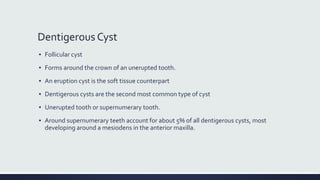
  • 15. Dentigerous Cyst ▪ Follicular cyst ▪ Forms around the crown of an unerupted tooth. ▪ An eruption cyst is the soft tissue counterpart ▪ Dentigerous cysts are the second most common type of cyst ▪ Unerupted tooth or supernumerary tooth. ▪ Around supernumerary teeth account for about 5% of all dentigerous cysts, most developing around a mesiodens in the anterior maxilla.
  • 16. Dentigerous Cyst ▪ Radiographic Features ▪ Location; Most commonly the mandibular or maxillary third molar or the maxillary canine. ▪ Diagnostic point is that this cyst attaches at the CEJ. ▪ Lateral Dentigerous Cyst; beside the crown instead of above the crown. ▪ Cysts related to maxillary third molars often grow into the maxillary antrum ▪ Cysts attached to the crown of mandibular molars may extend a considerable distance into the ramus. ▪ Well-DefinedCortex with Radiolucent internal structure ▪ Effects on surrounding Structure; Resorption/ Displacement
  • 17. Dentigerous Cyst ▪ Differential Diagnosis ▪ Hyperplastic Follicle; The size of the normal follicular space is 2 to 3 mm ▪ A cyst should be considered with any evidence of tooth displacement or considerable expansion of the involved bone ▪ Odontogenic Keratocyst; Does not expand the bone similar to Dentigerous, less likely to resorb teeth, and may attach further apically on the root ▪ Ameloblastic Fibroma ▪ Cystic Ameloblastoma ▪ SURGICAL REMOVAL
  • 23. Buccal Bifurcation Cyst ▪ Mandibular Infected Buccal Cyst ▪ Inflammatory /Paradental cyst ▪ Epithelial cell rests in the periodontal membrane of the buccal bifurcation of mandibular molars ▪ A common sign is the lack of or a delay in eruption of a mandibular first or second molar. ▪ On clinical examination the molar may be missing or the lingual cusp tips may be abnormally protruding through the mucosa, higher than the position of the buccal cusps. ▪ The first molar is involved more frequently than is the second molar. ▪ The teeth are always vital. ▪ The age of detection is younger, within the first two decades for a BBC rather than the third decade with a paradental cyst of the third molar.
  • 24. Buccal Bifurcation Cyst ▪ Radiographic Features ▪ Location. The mandibular first molar is the most common location followed by the second molar, occasionally is bilateral ▪ Periphery and Shape; Not readily apparent, and the lesion may be a very subtle radiolucent region superimposed over the image of the roots of the molar. ▪ In other cases the lesion has a circular shape with a well-defined cortical border. ▪ Internal Structure. The internal structure is radiolucent. ▪ Effects on Surrounding Structures. The most striking diagnostic characteristic of a BBC is the tipping of the involved molar ▪ The root tips are pushed into the lingual cortical plate of the mandible ▪ The occlusal surface is tipped toward the buccal aspect of the mandible
  • 25. Buccal Bifurcation Cyst ▪ Differential Diagnosis ▪ Periodontal Abscess ▪ Langerhans’ Cell Histiocytosis ▪ Dentigerous Cyst ▪ Management; Removal by conservative Curettage ▪ The involved molar should not be removed. ▪ BBCs do not recur.
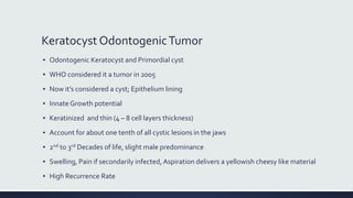
  • 27. Keratocyst OdontogenicTumor ▪ Odontogenic Keratocyst and Primordial cyst ▪ WHO considered it a tumor in 2005 ▪ Now it’s considered a cyst; Epithelium lining ▪ InnateGrowth potential ▪ Keratinized and thin (4 – 8 cell layers thickness) ▪ Account for about one tenth of all cystic lesions in the jaws ▪ 2nd to 3rd Decades of life, slight male predominance ▪ Swelling, Pain if secondarily infected, Aspiration delivers a yellowish cheesy like material ▪ High Recurrence Rate
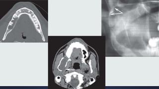
  • 28. Keratocyst OdontogenicTumor ▪ Radiographic Features ▪ Location; Posterior body of the mandible (90% occur posterior to the canines) and ramus (more than 50%) ▪ The epicenter is located superior to the inferior alveolar nerve canal. ▪ Periphery and Shape; cortical border unless they have become secondarily infected. ▪ Internal Structure; Most commonly radiolucent. ▪ Effects on Surrounding Structures; Grows along the internal aspect of the jaws, causing minimal expansion
  • 29. Keratocyst OdontogenicTumor ▪ Differential Diagnosis ▪ Dentigerous Cyst ▪ If several KOTs are found (which occurs in 4% to 5% of cases), these tumors may constitute part of a basal cell nevus syndrome. ▪ Management ▪ Enucleation and Curettage
  • 33. Basal Cell Nevus Syndrome ▪ Gorlin - Goltz Syndrome ▪ Nevoid Basal Cell Carcinoma Syndrome ▪ Multiple Keratocysts ▪ Multiple Nevoid Basal Cell Carcinomas of the skin, ▪ Skeletal abnormalities; bifid rib (most common), agenesis, deformity, and synostosis of the ribs, kyphoscoliosis, vertebral fusion, polydactyly, shortening of the metacarpals, temporal and temporo-parietal bossing, minor hyper-telorism ▪ Central nervous system abnormalities ▪ Eye abnormalities
  • 34. Gorlin-Goltz Syndrome ▪ Radiographic Features ▪ Location; the same as that of solitary KOTs, ▪ The multiple KOTs may develop bilaterally and can vary in size from 1 mm to several centimeters in diameter ▪ Other Radiographic Features ▪ Radiopaque line of the calcified falx cerebri may be prominent on the PA skull projection. ▪ Differential Diagnosis; Cherubism, Multiple Myeloma ▪ Management; More Aggressive
  • 36. Lateral Periodontal Cyst ▪ Arise from epithelial rests in periodontium lateral to the tooth root. ▪ Usually is unicystic ▪ Botryoid odontogenic cysts; Cluster of small Cysts ▪ Clinical Features ▪ Asymptomatic and less than 1 cm in diameter. ▪ No apparent sexual predilection, and the age distribution extends from the second to the ninth decades ▪ If these cysts become secondarily infected, they will mimic a lateral periodontal abscess.
  • 37. Lateral Periodontal Cyst ▪ Radiographic Features ▪ Location. A total of 50% to 75% of these cysts develop in the mandible, mostly in a region extending from the lateral incisor to the second premolar; Maxilla, between the lateral incisor and the cuspid. ▪ Periphery and Shape; Well-defined radiolucency with a prominent cortical boundary; Multilocular ▪ Effects on Surrounding Structures. ▪ Small cysts may efface the lamina dura of the adjacent root. ▪ Large cysts can displace adjacent teeth and cause expansion. ▪ Differential Diagnosis; Mental Foramen, Small Neurofibroma, Lateral RadicularCyst, Small Ameloblastoma ▪ Management
  • 39. Glandular OdontogenicTumor ▪ Sialo-odontogenic cyst ▪ Arising from odontogenic epithelium with a spectrum of characteristics including salivary gland features such as mucus-producing cells. ▪ Slight female predominance, has an aggressive behavior and a tendency to recur after surgery. ▪ Radiographic Features ▪ Location; More commonly in the mandible and most often in the anterior mandible and in the maxilla, commonly the globule-maxillary region. ▪ Periphery and Shape. There is usually a cortical boundary that may be smooth or scalloped. ▪ Internal Structure; Unilocular and multilocular appearances ▪ Effects on Surrounding Structures. Perforation through the cortex and Displacement of teeth is a common feature. ▪ Differential Diagnosis; Ameloblastoma, KOT ▪ AggressiveTreatment
  • 42. CalcifyingCystic OdontogenicTumor ▪ Calcifying odontogenic cyst ▪ Calcifying epithelial odontogenic cyst ▪ Gorlin cyst ▪ TheWorld Health Organization now categorizes this entity as a tumor. ▪ Uncommon, slow-growing, Painless, benign lesions. ▪ 2 peaks; First at 10 to 19 years of age and during the seventh decade ▪ Aspiration often yields a viscous, granular, yellow fluid. ▪ Most (75%) occur anterior to the first molar, especially associated with cuspids and incisors, where the cyst sometimes manifests as a pericoronal radiolucency. ▪ Occasionally (20% to 50% of cases) this tumor is associated with a tooth (most commonly a cuspid) and impedes its eruption.
  • 43. CCOT ▪ Differential Diagnosis; Dentigerous Cyst ▪ Adenomatoid OdontogenicTumor ▪ Ameloblastic Fibro-Odontoma ▪ Calcifying Epithelial OdontogenicTumor ▪ Management; Enucleation and curettage ▪ Periodic Follow-up
  • 45. Nasopalatine Duct Cyst ▪ IncisiveCanalCyst ▪ Median PalatineCyst; Posteriorly involving the hard palate ▪ 10% of cysts in the jaws ▪ Remnants of the Nasopalatine Duct cells ▪ Males * 3 times ▪ Small, well-defined swelling just posterior to the palatine papilla. ▪ Palatal to Incisors area ▪ Heart-Shaped Radiolucency (> 6 mm)
  • 48. Nasolabial/ Naso-Alveolar Cyst ▪ Small, it may produce a very subtle, unilateral swelling of the nasolabial fold and may elicit pain or discomfort. ▪ When large, it may bulge into the floor of the nasal cavity, causing some obstruction, flaring of the alae, distortion of the nostrils, and fullness of the upper lip. ▪ The age of detection ranges from 12 to 75 years, with a mean age of 44 years. ▪ About 75% of these lesions occur in females. ▪ Adjacent to the alveolar process above the apices of the incisors. ▪ MRI; SoftTissue ▪ Surgical Removal; Intra-Oral Approach
  • 50. Dermoid Cysts ▪ Cystic form of a teratoma thought to be derived from trapped embryonic cells that are toti-potential. ▪ The resulting cysts are lined with epidermis and cutaneous appendages and filled with keratin or sebaceous material (and in rare cases with bone, teeth, muscle, or hair, in which case they are properly called teratomas). ▪ Clinical Features ▪ May develop at any time from birth, but they usually become clinically apparent between 12 and 25 years ▪ The swelling, which is slow and painless, can grow to several centimeters in diameter, ▪ If located in the neck or tongue, it may interfere with breathing, speaking, and eating.
  • 51. Dermoid Cyst ▪ Location; About 10% or fewer arise in the head and neck ▪ Only 1% to 2% develop in the oral cavity; Of these, about 25% occur in the floor of the mouth and on the tongue. ▪ Periphery and Shape. Well-defined by more radiopaque soft tissue of this cyst compared with surrounding soft tissue, as seen inCT scans. ▪ Internal Structure; Radiolucent on conventional radiographs. ▪ Differential Diagnosis ▪ Ranula (unilateral or bilateral blockage ofWharton’s ducts),Thyroglossal duct cysts, cystic Hygromas, Branchial cleft cysts, cellulitis, tumors (lipoma and liposarcoma), and normal fat masses in the submental area. ▪ Management; Do not recur after surgical removal.
  • 53. Simple Bone Cyst ▪ Traumatic bone cyst, hemorrhagic bone cyst, extravasation cyst, progressive bone cavity, solitary bone cyst, and unicameral bone cyst ▪ Cavity within bone that is lined with connective tissue. ▪ It may be empty, or may contain fluid. ▪ However, because it has no epithelial lining, it is not a true cyst. ▪ The lesion shows a male predominance of approximately 2 : 1. ▪ Multiple SBCs can develop, especially when the disorder occurs with cemento-osseous dysplasia. ▪ The occurrence of SBCs in cemento-osseous dysplasia is seen in an older population, with a mean age of 42 years, and with a female predominance of 4 : 1. SBCs are asymptomatic in most cases. ▪ Aspiration usually produces only a few milliliters of straw-colored or serous-anguineous fluid.
  • 54. Simple Bone Cyst ▪ Location. Almost all SBCs are found in the mandible ▪ Periphery and Shape; May vary from a well-defined, delicate cortex to an ill-defined border that blends into the surrounding bone. ▪ Internal Structure; totally radiolucent ▪ Differential Diagnosis ▪ KOT ▪ Fibrous Dysplasia ▪ Management; Marsupialization and Curettage
  • 59. References ▪ Chapter 21: Cysts and Cyst-Like Lesions of the Jaws