CA Rectum
Presented By:
Dr. MONSIF IQBAL
Dept. of Surgery
POF Hospital, Wah Cantt, Pakistan
CASE
PRESENTATION
PATIENT’s PROFILE:
• Name: XYZ
• Age: 60 yrs.
• Sex: Male
• Address : Islamabad.
• D.O.A: 04-09-2013
• M.O.A: OPD
PRESENTING COMPLAINTS
• Constipation 1.5 years
• Bleeding PR 6 months
• Diagnosed case of CA Rectum
Past History
Sigmoidoscopy done in POF Hospital, Adenocarcinoma Rectum
(March--April 2013)
Past History Cont.
CT Scan Abdomen+Pelvis was done (April 2013)
Past History Cont.
• Neo-adjuvant Chemotheray and radiotherapy in
PIMS and Nescom Hospital
PHYSICAL EXAMINATION:
On Examination
▫ Pulse: 92/min
▫ B.P: 135/70 mm of Hg
▫ Oxygen Sat: 99%
On Abdominal Examination
 Distended
 Non tender
INVESTIGATIONS
1. Blood CP:
• Hb ---- 11.3 gm/dl
• TLC ---- 11.6x10³
/ul
• PLT ---- 434x103
/ul
2. ALT: 57
3. RFTs: Urea: 34, Creatinine 0.75
sodium : 139
Potassium : 4.0
Chloride : 101
4. CEA : 125
CT Scan Abdomen+Pelvis was done (Sep 2013)
Management
• IV fluids
• IV antibiotics and Analgesics started
• Councelled for APR
• Planned APR
Rectal Cancer
Rectal Cancer
Rectal Cancer
Rectal Cancer
Rectal Cancer
Rectal Cancer
Rectal Cancer
CA Rectum
Clinical Anatomy
• 12-15 cm from anal verge.
• Diameter
▫ 4 cm (upper part)
▫ Dilated (lower part)
• Posterior part of the
lesser pelvis and in front
of lower three pieces of
sacrum and the coccyx
• Begins at the
rectosigmoid junction, at
level of third sacral
vertebra
• Ends at the anorectal
junction, 2-3 cm in front
of and a little below the
coccyx
• Divided into 3 parts
• Upper third
• Middle third
• Lower third
• 3 distinct intraluminal
curves ( Valves of
Houston)
• Superior 1/3rd of the rectum
▫ Covered by peritoneum on the
anterior and lateral surfaces
• Middle 1/3rd of the rectum
▫ Covered by peritoneum on the
anterior surface
• Inferior 1/3rd of the rectum
▫ Devoid of peritoneum
▫ Close proximity to adjacent
structure including boney pelvis.
Peritoneal Relations
Arterial supply
• Superior rectal A – fr. IMA; supplies
upper rectum
• Middle rectal A- fr. Internal iliac A.
(supplies middle rectum)
• Inferior rectal A- fr. Internal pudendal A.
supplies lower rectum
Venous drainage
▫ Superior rectal V- upper & middle third
rectum
▫ Middle rectal V- lower rectum and upper
anal canal
▫ Inferior rectal vein- lower anal canal
Innervations
• Sympathetic: L1-L3, Hypogastric
nerve
• ParaSympathetic: S2-S4
Lymphatic drainage
• Upper and middle rectum
▫ Pararectal lymph nodes,
located directly on the muscle
layer of the rectum
▫ Inferior mesenteric lymph
nodes, via the nodes along the
superior rectal vessels
• Lower rectum
▫ Sacral group of lymph nodes or
Internal iliac lymph nodes
• Below the dentate line
▫ Inguinal nodes and external iliac
chain
Epidemiology
• Colorectal caner is the third most frequently diagnosed cancer
in the US men and women.
• 108,070 new cases of colon cancer and 40,740 new cases of
rectal cancer in the US in 2008. Combined mortality for
colorectal cancer 49,960 in 2008.
• Worldwide approx. 1 million new cases p.a. are diagnosed,
with 529,000 deaths.
• Incidence rate in India is quite low about 2 to 8 per 100,000
• Incidence in Wah Cantt, Taxilla and surrounding Sub-urban
areas is 55---60 cases per year.
• Median age- 7th
decade but can occur any time in adulthood.
• Cecum 14 %
• Ascending colon 10 %
• Transverse colon 12 %
• Descending colon 7 %
• Sigmoid colon 25 %
• Rectosigmoid junct 0.9 %
• Rectum 23 %
 Etiological agents
 Environmental & dietary factors
 Chemical carcinogenesis.
 Associated risk factors
 Male Gender
 Family history of colorectal cancer
 Personal history of colorectal cancer, ovary, endometrial, breast
 Excessive BMI
 Processed meat intake
 Excessive alcohol intake
 Low folate consumption
 Neoplastic polyps.
 Hereditary Conditions (FAP, HNPCC)
Clinical Presentations
• Symptoms
▫ Asymptomatic
▫ Change in bowel habit (diarrhoea, constipation, narrow stool,
incomplete evacuation, tenesmus).
▫ Blood PR.
▫ Abdominal discomfort (pain, fullness, cramps, bloating,
vomiting).
▫ Weight loss, tiredness.
• Acute Presentations
▫ Intestinal obstruction.
▫ Perforation.
▫ Massive bleeding.
• Signs
▫ Pallor
▫ Abdominal mass
▫ PR mass
▫ Jaundice
▫ Nodular liver
▫ Ascites
Pathological features
WHO Classification
• Adenocarcinoma in situ
• Adenocarcinoma
• Mucinous (colloid) adenocarcinoma (>50% mucinous)
• Signet ring cell carcinoma (>50% signet ring cells)
• Squamous cell (epidermoid) carcinoma
• Adenosquamous carcinoma
• Small-cell (oat cell) carcinoma
• Medullary carcinoma
• Undifferentiated Carcinoma
Dukes classification-
Dukes A: Invasion into but not through the bowel wall.
Dukes B: Invasion through the bowel wall but not involving lymph
nodes.
Dukes C: Involvement of lymph nodes
Dukes D: Widespread metastases
Modified astler coller classification-
Stage A : Limited to mucosa.
Stage B1 : Extending into muscularis propria but not penetrating
through it; nodes not involved.
Stage B2 : Penetrating through muscularis propria; nodes not
involved
Stage C1 : Extending into muscularis propria but not penetrating
through it. Nodes involved
Stage C2 : Penetrating through muscularis propria. Nodes
involved
Stage D: Distant metastatic spread
Tis TTis T11 TT22 TT33 TT44
MucosaMucosa
Muscularis mucosaeMuscularis mucosae
SubmucosaSubmucosa
Muscularis propriaMuscularis propria
SubserosaSubserosa
SerosaSerosa
TNM Classification
TX Primary tumor cannot be assessed
T0 No evidence of primary tumor
Tis Carcinoma in situ: intraepithelial or invasion of lamina propria
T1 Tumor invades submucosa
T2 Tumor invades muscularis propria
T3 Tumor invades through the muscularis propria into pericolorectal tissues
T4a Tumor penetrates to the surface of the visceral peritoneum
T4b Tumor directly invades or is adherent to other organs or structures
TNM Classification
Stage Grouping
Prognostic factors
 Good prognostic
factors
 Old age
 Gender(F>M)
 Asymptomatic pts
 Polypoidal lesions
 Diploid
 Poor prognostic
factors
 Obstruction
 Perforation
 Ulcerative lesion
 Adjacent structures
involvement
 Positive margins
 LVSI
 Signet cell carcinoma
 High CEA
 Tethered and fixed
cancer
Stage and Prognosis
Stage 5-year Survival (%)
0,1 Tis,T1;No;Mo > 90
I T2;No;Mo 80-85
II T3-4;No;Mo 70-75
III T2;N1-3;Mo 70-75
III T3;N1-3;Mo 50-65
III T4;N1-2;Mo 25-45
IV M1 <3
Diagnostic Workup
• History—including family history of colorectal cancer
or polyps
• Physical examinations including DRE and complete
pelvic examination in women: size, location, ulceration,
mobile vs. tethered vs. fixed, distance from anal verge
and sphincter functions.
• Proctoscopy—including assessment of mobility,
minimum diameter of the lumen, and distance from the
anal verge
• Biopsy of the primary tumor
Colonoscopy or barium enema
Figure: Carcinoma of the rectum. Double-
contrast barium enema shows a long
segment of concentric luminal narrowing
(arrows) along the rectum with minimal
irregularity of the mucosal surface.
To evaluate remainder of large bowel to rule out synchronous
tumor or presence of polyp syndrome.
Transrectal ultrasound –EUS
• use for clinical staging.
• 80-95% accurate in tumor staging
• 70-75% accurate in mesorectal lymph
node staging
• Very good at demonstrating layers of
rectal wall
• Use is limited to lesion < 14 cm from
anus, not applicable for upper rectum,
for stenosing tumor
• Very useful in determining extension of
disease into anal canal (clinical
important for planning sphincter
preserving surgery)
Figure. Endorectal
ultrasound of a T3 tumor of
the rectum, extension
through the muscularis
propria, and into perirectal
fat.
CT scan
• Part of routine workup of patients
• Useful in identifying enlarged pelvic lymph-nodes and
metastasis outside the pelvis than the extent or stage of
primary tumor
• Limited utility in small primary cancer
• Sensitivity 50-80%
• Specificity 30-80%
• Ability to detect pelvic and para-aortic lymph nodes is
higher than peri-rectal lymph nodes.
Magnetic Resonance Imaging (MRI)
• Greater accuracy in defining extent of rectal cancer
extension and also location & stage of tumor
• Also helpful in lateral extension of disease, critical in
predicting circumferential margin for surgical
excision.
• Different approaches (body coils, endorectal MRI &
phased array technique)
Figure: Mucinous adenocarcinoma of
the rectum. T2-weighted MRI shows high
signal intensity (arrowheads) of the
cancer lesion in right anterolateral side
of the rectal wall.
Figure: Normal rectal and perirectal
anatomy on high-resolution T2-weighted
MRI. Rectal mucosa (M), submucosa
(SM), and muscularis propria (PM) are
well discriminated. Mesorectal fascia
appears as a thin, low-signal-intensity
structure (arrowheads) and fuses with the
remnant of urogenital septum making
Denonvilliers fascia (arrows).
PET
• Shows promise as the most sensitive study
for the detection of metastatic disease in
the liver and elsewhere.
• Sensitivity of 97% and specificity of 76% in
evaluating for recurrent colorectal cancer.
cancer
rectum
prostate pubic bone
bladder
Small bowel
• CEA: High CEA levels associated with poorer
survival
• Routine investigation
▫ Complete blood count, RFT, LFT
▫ Chest X-ray
Rectal Cancer
Surgery
• Surgery is the mainstay of treatment of RC
• Local recurrence after conventional surgery:
▫ 20%-50% (average of 35%)**
• Radiotherapy significantly reduces the number
of local recurrences
Types of Surgery
• Local excision- reserved for superficially
invasive (T1) tumors with low likelihood of LN
metastases
• Should be considered a total biopsy, with further
treatment based on pathology
• With unfavorable pathology patient should undergo
total mesorectal excision with or without sphincter-
preservation:
▫ positive margin (or <2 mm), lymphovascular invasion,
▫ poorly differentiated tumors, T2 lesion
• Low Anterior Resection - for tumors in upper/mid
rectum; allows preservation of anal sphincter
• Abdominoperineal resection
▫ for tumors of distal rectum with distal edge up to 6 cm from anal
verge
▫ associated with permanent colostomy and high incidence of
sexual and genitourinary dysfunction
Complications of Surgery
• Bleeding
• Infection
• Anastomotic Leakage
• Blood clots
• Anesthetic Risks
Rectal Cancer
Purpose of Radio(chemo)therapy in Rectal
Cancer
• To lower local failure rates and improve survival in
resectable cancers
• to allow surgery in primarly inoperable cancers
• to facilitate a sphincter-preserving procedure
• to cure patients without surgery: very small cancer
or very high surgical risk
 5Fu
 Leucovorin
 Oxaliplatin
 Irinotecan
 Bevacizumab
 cetuximab
Combinations
 FOLFOX
 FOLFIRI
 Leucovorin/5FU
 Capecitabine
 Bevacizumab in
combination with the
above regimens.
Chemotherapy agents
Radiotherapy
• Preoperative radiotherapy
▫ Short course: 25 Gy in 5 daily fractions of 5 Gy given in 1 week.
▫ Long course
Phase 1
45 Gy in 25 daily fractions of 1.8 Gy given in 5 weeks.
Phase 2 (optional)
5.4–9 Gy in 3–5 daily fractions of 1.8 Gy
• Postoperative radiotherapy
Phase 1
45 Gy in 25 daily fractions of 1.8 Gy given in 5 weeks.
Phase 2 (optional)
5.4–9 Gy in 3–5 daily fractions of 1.8 Gy.
• Palliative radiotherapy
Clinical Trials
Pre-op RT vs. surgery alone
Swedish Rectal Cancer Trial(NEJM 1997;336:980 ): 1168 patients
randomised to 25 Gy (5x5) PRT or no RT.
Surgery alone Preop. RT
Rate of local recurrence 27% 11% p<0.001
5-year overall survival 48% 58% p=0.004
Dutch Colorectal Cancer Group (Kapiteijn E. NEJM 2001;345:638):
1861 patients randomised TME vs PRT+TME
TME PRT+TME
Recurrence rate 2.4% 8.2%
OS ns ns
Pre-op vs. post-op Chemo RT
Randomized trial of the German Rectal Cancer study
Group (Sauer R et al. N Engl J Med 2004;351:1731-40):
▫ cT3 or cT4 or node-positive rectal cancer
▫ 50,4 Gy (1.8 Gy per day)
▫ 5-FU: 1000 mg/m2
per day (d1-5) during 1. and 5. week
Preop CRT Postop CRT
Patients N=415 N=384
5 y. OS 76% 74% p=0.8
5 y. local relapse 6% 13% p=0.006
G3,4 toxic effects 27% 40% p=0.001
• Increase in sphincter-preserving surgery with preop Th.
Thank You !!

More Related Content

PPT
Carcinoma rectum
PPTX
Rectal cancer alex
PPTX
Ca rectum
PPTX
RECTAL CANCER adesiyakan
PPTX
Carcinoma rectum
PPTX
Ca rectum Management seminar 2019
PPTX
CARCINOMA RECTUM MANAGEMENT
PPTX
Rectal Carcinoma
Carcinoma rectum
Rectal cancer alex
Ca rectum
RECTAL CANCER adesiyakan
Carcinoma rectum
Ca rectum Management seminar 2019
CARCINOMA RECTUM MANAGEMENT
Rectal Carcinoma

What's hot (20)

PPTX
Clinical examination of abdominal lump
PPTX
Benign breast disease
PPTX
testicular tumors
PPTX
Carcinoma colon
PPTX
Carcinoma Rectum
PPTX
Benign breast disease and its management
PPTX
Ca rectum
PPTX
Splenic trauma
PPTX
Gastric Cancer / Carcinoma management
PPT
Carcinoma rectum (Rectal Cancer)
PPTX
familial adenomatous polyposis
PPTX
Carcinoma Of Prostate and its management
PPT
Retroperitoneal mass
PPTX
Cholangiocarcinoma
PPT
Carcinoma oesophagus
PPT
Anal cancer ppt
PPTX
SPLENIC INJURY.pptx
PPT
Lower GI - Bleed
PPTX
Penile carcinoma
PPTX
Post gastrectomy syndrome
Clinical examination of abdominal lump
Benign breast disease
testicular tumors
Carcinoma colon
Carcinoma Rectum
Benign breast disease and its management
Ca rectum
Splenic trauma
Gastric Cancer / Carcinoma management
Carcinoma rectum (Rectal Cancer)
familial adenomatous polyposis
Carcinoma Of Prostate and its management
Retroperitoneal mass
Cholangiocarcinoma
Carcinoma oesophagus
Anal cancer ppt
SPLENIC INJURY.pptx
Lower GI - Bleed
Penile carcinoma
Post gastrectomy syndrome

Similar to Rectal Cancer (20)

PPT
carcinomarectum-111113085726-phpapp01 (1).ppt
PPT
Carcinomarectum 111113085726-phpapp01
PPTX
Ca rectum premanagement
PPTX
Carcinoma rectum-radiotherapy perspective
PPTX
Rectal cancer
PPTX
PREMANAGEMENT & MANAGEMENT OF Ca RECTUM.pptx
PPT
Carcinoma.ppt
PPTX
Rectal cancer By Dr Efrem Ayalew Wossen
PPTX
Rectal carcinoma approach
PPTX
Carcinoma rectum the complete aproach to how to investigate and treat a case ...
PPTX
ca rectum new2.pptx
PPTX
Colorectal Carcenoma - Copy by undergraduate .pptx
PPTX
Management of Rectal cancer.pptx
PPTX
Ca. Rectum.pptx
PPTX
Carcinoma rectum
PPTX
carcinoma rectum and anal canal.pptx
PDF
COLORECTAL CANCER.pdf
PPTX
Carcinoma Rectum by Dr.K.AmrithaAnilkumar
PPTX
Locally Advanced Rectal Cancer
PPTX
approach for rectal carcinoma and management
carcinomarectum-111113085726-phpapp01 (1).ppt
Carcinomarectum 111113085726-phpapp01
Ca rectum premanagement
Carcinoma rectum-radiotherapy perspective
Rectal cancer
PREMANAGEMENT & MANAGEMENT OF Ca RECTUM.pptx
Carcinoma.ppt
Rectal cancer By Dr Efrem Ayalew Wossen
Rectal carcinoma approach
Carcinoma rectum the complete aproach to how to investigate and treat a case ...
ca rectum new2.pptx
Colorectal Carcenoma - Copy by undergraduate .pptx
Management of Rectal cancer.pptx
Ca. Rectum.pptx
Carcinoma rectum
carcinoma rectum and anal canal.pptx
COLORECTAL CANCER.pdf
Carcinoma Rectum by Dr.K.AmrithaAnilkumar
Locally Advanced Rectal Cancer
approach for rectal carcinoma and management

More from Monsif Iqbal (8)

PPTX
Intravenous Fluids in Surgical Practice
PPT
Hospital aquired infections
PPT
Surgical nutrition
PPT
Polytrauma
PPT
Esophageal Cancer
PPT
Liver trauma
PPT
Subdural Hematoma
PPTX
Spinal Tuberculosis by Dr. Monsif Iqbal
Intravenous Fluids in Surgical Practice
Hospital aquired infections
Surgical nutrition
Polytrauma
Esophageal Cancer
Liver trauma
Subdural Hematoma
Spinal Tuberculosis by Dr. Monsif Iqbal

Recently uploaded (20)

PPTX
Right Lateral Medullary Syndrome (1).pptx
PPTX
SlideEgg_100085- World Mental Health Day.pptx
PPTX
PDF
Joint Commission EBPCD24_samplepages.pdf
PPTX
Emotional Well Being & Conflict Resolution_VKV.pptx
PPTX
Medical Legal issues in Psychiatry Final.pptx
PDF
CSF rhinorrhea its cause management .pptx
PPTX
Oncological Emergencies in hospital setting
PDF
Dental Implants Review : A detailed Review
PPT
FRACTURE CLASSIFICATION AND MANAGEMENT..
PDF
CASE PRESENTATION1.pdf bipolar disorder in which both mania and depression h...
PPTX
Common Bacterial infections-converted_64bcdc4f77a3b7b90bdeb611f66c6ddd.pptx
PDF
Cellular Respiration-BIOLOGEYCHEMESTRY'S
PDF
Indonesian Healthtech Innovation_11Sep2019_Industry_Geraldine Seow_1.pdf
PPTX
CLASS III MALOCCLUSION IN ORTHODONTICS
PPTX
The Process of Infection by Windy Mesolas-Luzon.pptx
PPT
NEPHROTIC SYNDROME POWER POINT PRESENTATION
PPT
toxicosis in pregnancy preeclampcia eclampcia.ppt
PPTX
health care concerns.pptx by hemant kumari
PDF
Chapter 8. HHS Facility Design and Construction _ HHS.gov.pdf
Right Lateral Medullary Syndrome (1).pptx
SlideEgg_100085- World Mental Health Day.pptx
Joint Commission EBPCD24_samplepages.pdf
Emotional Well Being & Conflict Resolution_VKV.pptx
Medical Legal issues in Psychiatry Final.pptx
CSF rhinorrhea its cause management .pptx
Oncological Emergencies in hospital setting
Dental Implants Review : A detailed Review
FRACTURE CLASSIFICATION AND MANAGEMENT..
CASE PRESENTATION1.pdf bipolar disorder in which both mania and depression h...
Common Bacterial infections-converted_64bcdc4f77a3b7b90bdeb611f66c6ddd.pptx
Cellular Respiration-BIOLOGEYCHEMESTRY'S
Indonesian Healthtech Innovation_11Sep2019_Industry_Geraldine Seow_1.pdf
CLASS III MALOCCLUSION IN ORTHODONTICS
The Process of Infection by Windy Mesolas-Luzon.pptx
NEPHROTIC SYNDROME POWER POINT PRESENTATION
toxicosis in pregnancy preeclampcia eclampcia.ppt
health care concerns.pptx by hemant kumari
Chapter 8. HHS Facility Design and Construction _ HHS.gov.pdf

Rectal Cancer

  • 1. CA Rectum Presented By: Dr. MONSIF IQBAL Dept. of Surgery POF Hospital, Wah Cantt, Pakistan
  • 3. PATIENT’s PROFILE: • Name: XYZ • Age: 60 yrs. • Sex: Male • Address : Islamabad. • D.O.A: 04-09-2013 • M.O.A: OPD
  • 4. PRESENTING COMPLAINTS • Constipation 1.5 years • Bleeding PR 6 months • Diagnosed case of CA Rectum
  • 5. Past History Sigmoidoscopy done in POF Hospital, Adenocarcinoma Rectum (March--April 2013)
  • 6. Past History Cont. CT Scan Abdomen+Pelvis was done (April 2013)
  • 7. Past History Cont. • Neo-adjuvant Chemotheray and radiotherapy in PIMS and Nescom Hospital
  • 8. PHYSICAL EXAMINATION: On Examination ▫ Pulse: 92/min ▫ B.P: 135/70 mm of Hg ▫ Oxygen Sat: 99% On Abdominal Examination  Distended  Non tender
  • 9. INVESTIGATIONS 1. Blood CP: • Hb ---- 11.3 gm/dl • TLC ---- 11.6x10³ /ul • PLT ---- 434x103 /ul 2. ALT: 57 3. RFTs: Urea: 34, Creatinine 0.75 sodium : 139 Potassium : 4.0 Chloride : 101 4. CEA : 125
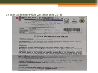
  • 10. CT Scan Abdomen+Pelvis was done (Sep 2013)
  • 11. Management • IV fluids • IV antibiotics and Analgesics started • Councelled for APR • Planned APR
  • 20. Clinical Anatomy • 12-15 cm from anal verge. • Diameter ▫ 4 cm (upper part) ▫ Dilated (lower part) • Posterior part of the lesser pelvis and in front of lower three pieces of sacrum and the coccyx • Begins at the rectosigmoid junction, at level of third sacral vertebra • Ends at the anorectal junction, 2-3 cm in front of and a little below the coccyx
  • 21. • Divided into 3 parts • Upper third • Middle third • Lower third • 3 distinct intraluminal curves ( Valves of Houston)
  • 22. • Superior 1/3rd of the rectum ▫ Covered by peritoneum on the anterior and lateral surfaces • Middle 1/3rd of the rectum ▫ Covered by peritoneum on the anterior surface • Inferior 1/3rd of the rectum ▫ Devoid of peritoneum ▫ Close proximity to adjacent structure including boney pelvis. Peritoneal Relations
  • 23. Arterial supply • Superior rectal A – fr. IMA; supplies upper rectum • Middle rectal A- fr. Internal iliac A. (supplies middle rectum) • Inferior rectal A- fr. Internal pudendal A. supplies lower rectum Venous drainage ▫ Superior rectal V- upper & middle third rectum ▫ Middle rectal V- lower rectum and upper anal canal ▫ Inferior rectal vein- lower anal canal Innervations • Sympathetic: L1-L3, Hypogastric nerve • ParaSympathetic: S2-S4
  • 24. Lymphatic drainage • Upper and middle rectum ▫ Pararectal lymph nodes, located directly on the muscle layer of the rectum ▫ Inferior mesenteric lymph nodes, via the nodes along the superior rectal vessels • Lower rectum ▫ Sacral group of lymph nodes or Internal iliac lymph nodes • Below the dentate line ▫ Inguinal nodes and external iliac chain
  • 25. Epidemiology • Colorectal caner is the third most frequently diagnosed cancer in the US men and women. • 108,070 new cases of colon cancer and 40,740 new cases of rectal cancer in the US in 2008. Combined mortality for colorectal cancer 49,960 in 2008. • Worldwide approx. 1 million new cases p.a. are diagnosed, with 529,000 deaths. • Incidence rate in India is quite low about 2 to 8 per 100,000 • Incidence in Wah Cantt, Taxilla and surrounding Sub-urban areas is 55---60 cases per year. • Median age- 7th decade but can occur any time in adulthood.
  • 26. • Cecum 14 % • Ascending colon 10 % • Transverse colon 12 % • Descending colon 7 % • Sigmoid colon 25 % • Rectosigmoid junct 0.9 % • Rectum 23 %
  • 27.  Etiological agents  Environmental & dietary factors  Chemical carcinogenesis.  Associated risk factors  Male Gender  Family history of colorectal cancer  Personal history of colorectal cancer, ovary, endometrial, breast  Excessive BMI  Processed meat intake  Excessive alcohol intake  Low folate consumption  Neoplastic polyps.  Hereditary Conditions (FAP, HNPCC)
  • 28. Clinical Presentations • Symptoms ▫ Asymptomatic ▫ Change in bowel habit (diarrhoea, constipation, narrow stool, incomplete evacuation, tenesmus). ▫ Blood PR. ▫ Abdominal discomfort (pain, fullness, cramps, bloating, vomiting). ▫ Weight loss, tiredness. • Acute Presentations ▫ Intestinal obstruction. ▫ Perforation. ▫ Massive bleeding.
  • 29. • Signs ▫ Pallor ▫ Abdominal mass ▫ PR mass ▫ Jaundice ▫ Nodular liver ▫ Ascites
  • 30. Pathological features WHO Classification • Adenocarcinoma in situ • Adenocarcinoma • Mucinous (colloid) adenocarcinoma (>50% mucinous) • Signet ring cell carcinoma (>50% signet ring cells) • Squamous cell (epidermoid) carcinoma • Adenosquamous carcinoma • Small-cell (oat cell) carcinoma • Medullary carcinoma • Undifferentiated Carcinoma
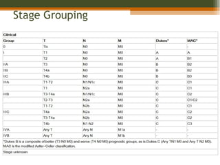
  • 31. Dukes classification- Dukes A: Invasion into but not through the bowel wall. Dukes B: Invasion through the bowel wall but not involving lymph nodes. Dukes C: Involvement of lymph nodes Dukes D: Widespread metastases Modified astler coller classification- Stage A : Limited to mucosa. Stage B1 : Extending into muscularis propria but not penetrating through it; nodes not involved. Stage B2 : Penetrating through muscularis propria; nodes not involved Stage C1 : Extending into muscularis propria but not penetrating through it. Nodes involved Stage C2 : Penetrating through muscularis propria. Nodes involved Stage D: Distant metastatic spread
  • 32. Tis TTis T11 TT22 TT33 TT44 MucosaMucosa Muscularis mucosaeMuscularis mucosae SubmucosaSubmucosa Muscularis propriaMuscularis propria SubserosaSubserosa SerosaSerosa TNM Classification TX Primary tumor cannot be assessed T0 No evidence of primary tumor Tis Carcinoma in situ: intraepithelial or invasion of lamina propria T1 Tumor invades submucosa T2 Tumor invades muscularis propria T3 Tumor invades through the muscularis propria into pericolorectal tissues T4a Tumor penetrates to the surface of the visceral peritoneum T4b Tumor directly invades or is adherent to other organs or structures
  • 35. Prognostic factors  Good prognostic factors  Old age  Gender(F>M)  Asymptomatic pts  Polypoidal lesions  Diploid  Poor prognostic factors  Obstruction  Perforation  Ulcerative lesion  Adjacent structures involvement  Positive margins  LVSI  Signet cell carcinoma  High CEA  Tethered and fixed cancer
  • 36. Stage and Prognosis Stage 5-year Survival (%) 0,1 Tis,T1;No;Mo > 90 I T2;No;Mo 80-85 II T3-4;No;Mo 70-75 III T2;N1-3;Mo 70-75 III T3;N1-3;Mo 50-65 III T4;N1-2;Mo 25-45 IV M1 <3
  • 37. Diagnostic Workup • History—including family history of colorectal cancer or polyps • Physical examinations including DRE and complete pelvic examination in women: size, location, ulceration, mobile vs. tethered vs. fixed, distance from anal verge and sphincter functions. • Proctoscopy—including assessment of mobility, minimum diameter of the lumen, and distance from the anal verge • Biopsy of the primary tumor
  • 38. Colonoscopy or barium enema Figure: Carcinoma of the rectum. Double- contrast barium enema shows a long segment of concentric luminal narrowing (arrows) along the rectum with minimal irregularity of the mucosal surface. To evaluate remainder of large bowel to rule out synchronous tumor or presence of polyp syndrome.
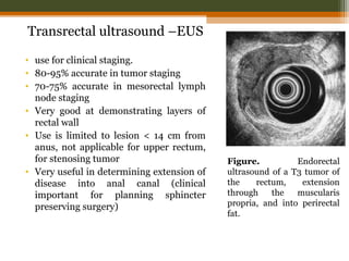
  • 39. Transrectal ultrasound –EUS • use for clinical staging. • 80-95% accurate in tumor staging • 70-75% accurate in mesorectal lymph node staging • Very good at demonstrating layers of rectal wall • Use is limited to lesion < 14 cm from anus, not applicable for upper rectum, for stenosing tumor • Very useful in determining extension of disease into anal canal (clinical important for planning sphincter preserving surgery) Figure. Endorectal ultrasound of a T3 tumor of the rectum, extension through the muscularis propria, and into perirectal fat.
  • 40. CT scan • Part of routine workup of patients • Useful in identifying enlarged pelvic lymph-nodes and metastasis outside the pelvis than the extent or stage of primary tumor • Limited utility in small primary cancer • Sensitivity 50-80% • Specificity 30-80% • Ability to detect pelvic and para-aortic lymph nodes is higher than peri-rectal lymph nodes.
  • 41. Magnetic Resonance Imaging (MRI) • Greater accuracy in defining extent of rectal cancer extension and also location & stage of tumor • Also helpful in lateral extension of disease, critical in predicting circumferential margin for surgical excision. • Different approaches (body coils, endorectal MRI & phased array technique)
  • 42. Figure: Mucinous adenocarcinoma of the rectum. T2-weighted MRI shows high signal intensity (arrowheads) of the cancer lesion in right anterolateral side of the rectal wall. Figure: Normal rectal and perirectal anatomy on high-resolution T2-weighted MRI. Rectal mucosa (M), submucosa (SM), and muscularis propria (PM) are well discriminated. Mesorectal fascia appears as a thin, low-signal-intensity structure (arrowheads) and fuses with the remnant of urogenital septum making Denonvilliers fascia (arrows).
  • 43. PET • Shows promise as the most sensitive study for the detection of metastatic disease in the liver and elsewhere. • Sensitivity of 97% and specificity of 76% in evaluating for recurrent colorectal cancer. cancer rectum prostate pubic bone bladder Small bowel
  • 44. • CEA: High CEA levels associated with poorer survival • Routine investigation ▫ Complete blood count, RFT, LFT ▫ Chest X-ray
  • 46. Surgery • Surgery is the mainstay of treatment of RC • Local recurrence after conventional surgery: ▫ 20%-50% (average of 35%)** • Radiotherapy significantly reduces the number of local recurrences
  • 47. Types of Surgery • Local excision- reserved for superficially invasive (T1) tumors with low likelihood of LN metastases • Should be considered a total biopsy, with further treatment based on pathology • With unfavorable pathology patient should undergo total mesorectal excision with or without sphincter- preservation: ▫ positive margin (or <2 mm), lymphovascular invasion, ▫ poorly differentiated tumors, T2 lesion
  • 48. • Low Anterior Resection - for tumors in upper/mid rectum; allows preservation of anal sphincter • Abdominoperineal resection ▫ for tumors of distal rectum with distal edge up to 6 cm from anal verge ▫ associated with permanent colostomy and high incidence of sexual and genitourinary dysfunction
  • 49. Complications of Surgery • Bleeding • Infection • Anastomotic Leakage • Blood clots • Anesthetic Risks
  • 51. Purpose of Radio(chemo)therapy in Rectal Cancer • To lower local failure rates and improve survival in resectable cancers • to allow surgery in primarly inoperable cancers • to facilitate a sphincter-preserving procedure • to cure patients without surgery: very small cancer or very high surgical risk
  • 52.  5Fu  Leucovorin  Oxaliplatin  Irinotecan  Bevacizumab  cetuximab Combinations  FOLFOX  FOLFIRI  Leucovorin/5FU  Capecitabine  Bevacizumab in combination with the above regimens. Chemotherapy agents
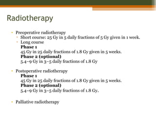
  • 53. Radiotherapy • Preoperative radiotherapy ▫ Short course: 25 Gy in 5 daily fractions of 5 Gy given in 1 week. ▫ Long course Phase 1 45 Gy in 25 daily fractions of 1.8 Gy given in 5 weeks. Phase 2 (optional) 5.4–9 Gy in 3–5 daily fractions of 1.8 Gy • Postoperative radiotherapy Phase 1 45 Gy in 25 daily fractions of 1.8 Gy given in 5 weeks. Phase 2 (optional) 5.4–9 Gy in 3–5 daily fractions of 1.8 Gy. • Palliative radiotherapy
  • 55. Pre-op RT vs. surgery alone Swedish Rectal Cancer Trial(NEJM 1997;336:980 ): 1168 patients randomised to 25 Gy (5x5) PRT or no RT. Surgery alone Preop. RT Rate of local recurrence 27% 11% p<0.001 5-year overall survival 48% 58% p=0.004 Dutch Colorectal Cancer Group (Kapiteijn E. NEJM 2001;345:638): 1861 patients randomised TME vs PRT+TME TME PRT+TME Recurrence rate 2.4% 8.2% OS ns ns
  • 56. Pre-op vs. post-op Chemo RT Randomized trial of the German Rectal Cancer study Group (Sauer R et al. N Engl J Med 2004;351:1731-40): ▫ cT3 or cT4 or node-positive rectal cancer ▫ 50,4 Gy (1.8 Gy per day) ▫ 5-FU: 1000 mg/m2 per day (d1-5) during 1. and 5. week Preop CRT Postop CRT Patients N=415 N=384 5 y. OS 76% 74% p=0.8 5 y. local relapse 6% 13% p=0.006 G3,4 toxic effects 27% 40% p=0.001 • Increase in sphincter-preserving surgery with preop Th.

Editor's Notes

  • #58: There are interesting things to be found when researching information on the internet to include in a presentation. The Colossal Colon is a replica of the human colon that is four feet wide. It was modeled from colonoscopy footage. It has traveled across the U.S. to inform the public about colon health. People can crawl through the colon or view through windows on the outside. It shows healthy colon tissue as well as diseased tissue including polyps and colon cancer. This picture was taken at a mall near the Creighton University Medical Center. It’s a fun way to spread information about colon health.