Shock
10.04.2020
Definition of shock
• Shock is a state in which there is inadequate blood flow to the tissues to
meet demand.
• Shock and hypotension often co-exist, BUT a normal blood pressure DOES
NOT exclude the diagnosis of shock
• Imbalance between tissue supply and demand →Anaerobic Metabolism
→Lactic Acid Production
• The acute circulatory failure if prolonged, results in irreversible organ
failure.
• Mortality is high without early diagnosis and treatment.
Department of Anaesthesia and Intensive Care, the Chinese Universityof Hong Kong July 2015
Medical Guidelines Medicins Sans Fronteires on Shock
Clinical features-- Signs common to most forms of
shock
– Pallor, mottled skin, cold extremities, sweating and thirst.
– Rapid and weak pulse often only detected on major arteries (femoral or
carotid).
– Low blood pressure (BP), narrow pulse pressure, BP sometimes
undetectable.
– Capillary refill time (CRT) > 3 seconds.
– Cyanosis, dyspnoea, tachypnoea are often present in varying degrees
depending on the mechanism.
– Consciousness usually maintained, but anxiety, confusion, agitation or
apathy are common.
– Oliguria or anuria.
Medical Guidelines Medicins Sans Fronteires on Shock
Different types of Shock
1. Hypovolaemic
2. Cardiogenic
3. Distributive
4. Obstructive
Department of Anaesthesia and Intensive Care, the Chinese Universityof Hong Kong
July 2015
Signs specific to the mechanism of shock --
Hypovolaemic shock
• The common signs of shock listed above are typical of hypovolaemic
shock.
• Do not underestimate hypovolaemia. Signs of shock may not become
evident until a 50% loss of blood volume in adults.
Medical Guidelines Medicins Sans Fronteires on Shock
Distributive Shock
• Caused by maldistribution of blood flow from peripheral
vasodilatation and decrease in systemic vascular resistance (SVR)
(decreased afterload)
• Sepsis
• Anaphylaxis
• Neurogenic
Signs specific to the mechanism of shock - Septic
shock
– High fever or hypothermia (< 36 °C), rigors, confusion
– BP may be initially maintained, but rapidly, same pattern as for
hypovolaemic shock.
Medical Guidelines Medicins Sans Fronteires on Shock
Signs specific to the mechanism of shock -
Anaphylactic shock
– Significant and sudden drop in BP
– Tachycardia
– Frequent cutaneous signs: rash, urticaria, angioedema
– Respiratory signs: dyspnoea, bronchospasm
Medical Guidelines Medicins Sans Fronteires on Shock
Signs specific to the mechanism of shock -
Cardiogenic shock
– Respiratory signs of left ventricular failure (acute pulmonary oedema)
are dominant: tachypnoea, crepitations on auscultation.
– Signs of right ventricular failure: raised jugular venous pressure,
hepatojugular reflux, sometimes alone, more often associated with
signs of left ventricular failure.
Medical Guidelines Medicins Sans Fronteires on Shock
Causes of
Cardiogenic
Shock
Hollenberg SM, Kavinsky CJ, ParrilloJE. Cardiogenic shock. Ann Intern Med. 1999;131(1):47-59.
Trauma Patients
Can Present with A
Combination of
Different Types of
Shock
Jeremy Richards et al. Diagnosis And Management Of Shock In The
EmergencyDepartment. EmergencyMedicine Practice 2014, 16:3
The aetiological diagnosis is oriented by:
– The context: trauma, insect bite, ongoing medical treatment, etc.
– The clinical examination:
• fever
• skin pinch consistent with dehydration
• thoracic pain from a myocardial infarction or pulmonary embolus
• abdominal pain or rigidity of the abdominal wall from peritonitis,
abdominal distension from intestinal obstruction
• blood in stools, vomiting blood in intestinal haemorrhage
• subcutaneous crepitations, likely anaerobic infection
Medical Guidelines Medicins Sans Fronteires on Shock
Characteristics of different types of shock
Type of shock JVP/CVP Cardiac output (CO) Systemic vascular
resistance (SVR)
Clinically
Hypovolemic ↓ ↓ ↑ Cold and shut down
Cardiogenic ↑ ↓ ↑ Cold and shut down
Distributive ↓ ↓/-/↑ ↓ Warm and dilated
Obstructive ↑ ↓ ↑ Cold and shut down
Department of Anaesthesia and Intensive Care, the Chinese Universityof Hong Kong
July 2015
Lab studies
• FBC
• RP
• LFT
• Amylase
• Cardiac biomarkers
• ABG
• Cultures
• Lactate
Jeremy Richards et al. Diagnosis And Management Of Shock In The
EmergencyDepartment. EmergencyMedicine Practice 2014, 16:3
Imaging
• USS
• Maximal inferior vena cava diameter was better able to differentiate a central venous
pressure <10mmHg from a central venous pressure >10mmHg
• Abdominal aortic aneurysm
• Echo
• Cardiac ejection fraction to determine etiology of shock – depressed in cardiogenic shock
• Cardiac tamponade
• Aortic dissection
• Pulmonary embolism
• CXR
• Heart size, presence/absence of pulmonary edema/infiltrates/effusion/ pneumothorax
• CT scan
• Only if USS is inconclusive
Jeremy Richards et al. Diagnosis And Management Of Shock In The
EmergencyDepartment. EmergencyMedicine Practice 2014, 16:3
Management of Shock (General)
• Definitive treatment for patients in shock depends on the specific
etiology; however, this may not be immediately clear on initial
presentation
• Thus initiation of therapy and patient stabilization may occur
simultaneously with evaluation.
• The goals in treating patients in shock are restoring adequate organ
perfusion and oxygen delivery while considering/treating the possible
cause(s) of shock
Jeremy Richards et al. Diagnosis And Management Of Shock In The
EmergencyDepartment. EmergencyMedicine Practice 2014, 16:3
Initial Stabilization
• Airway
• Breathing
• Circulation
Jeremy Richards et al. Diagnosis And Management Of Shock In The
EmergencyDepartment. EmergencyMedicine Practice 2014, 16:3
Management of Shock (General)
• Early recognition and prompt treatment of the underlying cause of
shock
• Ensure oxygenation and maintain perfusion
• Usually aim for:
• Mean Arterial Pressure (MAP) ≥ 65mmHg .…. formula MAP = SBP + 2(DBP)
3
• Urine output ≥ 0.5ml/kg/hr
Department of Anaesthesia and Intensive Care, the Chinese Universityof Hong Kong
July 2015
Treatment – Monitoring
• Cardiovascular monitoring
• Cardiac monitoring, BP cuffs automated
Jeremy Richards et al. Diagnosis And Management Of Shock In The
EmergencyDepartment. EmergencyMedicine Practice 2014, 16:3
Treatment -- Fluid resuscitation
• Only approximately 50% of hypotensive patients are volume responsive
• Passive leg raise can rapidly, noninvasively and easily as a reversible tool to
assess volume responsiveness
• Leg is raised from horizontal to vertical position and then assess volume
responsiveness
• Crystalloid is usually used for the initial treatment of undifferentiated shock
Jeremy Richards et al. Diagnosis And Management Of Shock In The
EmergencyDepartment. EmergencyMedicine Practice 2014, 16:3
Management -- Vasopressor
• Once patient is euvolemic but there is still ineffective oxygen delivery,
vasoactive medications are likely required
• Vasopressor medications used to support mean arterial pressure by
increasing systemic vascular resistance and/or cardiac output
• Noradrenaline is a strong alpha agonist with some beta 1 activity and is a
recommended initial choice for most categories of shock
• Metaanalysis of 11 trials showed there is a statistically significant increased
risk of death associated with dopamine. Thus should not be used as a first
line pressor agent
Jeremy Richards et al. Diagnosis And Management Of Shock In The
EmergencyDepartment. EmergencyMedicine Practice 2014, 16:3
Treating Shock – Vasopressors
Activity at Receptors
Agent α1 α2 𝛽1 𝛽2 Dopaminergic
Dobutamine + + ++++ ++ 0
Adrenaline ++++ ++++ ++++ +++ 0
Noradrenaline +++ +++ +++ +/++ 0
Restoring Blood Pressure
• Mean arterial pressure is the preferred blood pressure to target during resuscitation
• The recommended initial goal is 65 mm Hg. A higher goal of 80 to 85 mm Hg may help patients
with chronic hypertension, while a lower target may be better tolerated in patients with reduced
systolic function, older patients, and patients with end-stage liver disease
• After blood pressure falls below a critical threshold, tissue perfusion decreases linearly. That
critical threshold can vary between organ systems and individuals, and the target can later be
personalized based on global and regional perfusion as assessed with urine output, mental status,
or lactate clearance
• Decisions to titrate vasopressors to achieve mean arterial pressure goals should be balanced
against potential adverse effects, including arrhythmias, cardiovascular events, and ischemia
Siddharth Dugar et al Sepsis and septic shock: Guideline-based managementCLEVELAND CLINIC JOURNAL OF
MEDICINE VOLUME 87 • NUMBER 1 JANUARY 2020
Restoring Blood Pressure
• Norepinephrine is the first line vasopressor agent
• A second vasopressor is routinely added when norepinephrine doses
exceed 40 or 50 μg/min
• Epinephrine or vasopressin as second line vasopressor agent
Siddharth Dugar et al Sepsis and septic shock: Guideline-based managementCLEVELAND CLINIC JOURNAL OF
MEDICINE VOLUME 87 • NUMBER 1 JANUARY 2020
Hypovolaemic shock
- Due to inadequate circulating fluid volume
Causes:
- divided to haemorrhagic or non-haemorrhagic (major burns;
gastrointestinal losses: vomiting, fistulas; urinary losses: diabetes,
diabetes insipidus; evaporative losses with fever, abdominal surgery)
Management:
- fluid resuscitation
- haemorrhagic cause: transfusion of red cells and blood products.
- review source of bleeding and stop bleeding promptly
- use of hemostatic agent
Department of Anaesthesia and Intensive Care, the Chinese Universityof Hong Kong
July 2015
Cardiogenic shock
- due to cardiac pump failure resulting from myocardial or valvular failure
Causes:
- commonest: acute coronary syndrome
- other causes: arrhythmia, myocardial contusions post-trauma; myocarditis; acute valvular dysfunction;
cardiomyopathy
- echo is very useful in reviewing the cause and monitor the progress
Management:
- Acute Coronary Syndrome (ACS): reperfusion by fibrinolytics or Per Cutaneous Intervention (angiogram ±
angioplasty ± stenting)
- control arrhythmia: pharmacological, electrical: pacing/ cardioversion
- optimise preload by fluid: a trial of small bolus of fluid with close observation of CVP/BP trend
- inotropic support: augment myocardial contractility. Increasing diastolic blood pressure to increase coronary
perfusion pressure and flow
- afterload: vasodilator will cause further hypotension, use with caution in normotensive patients
- mechanical device: Intraaortic Ballon Pump (IABP), Extracorporeal Membrane Oxygenation (ECMO)
Department of Anaesthesia and Intensive Care, the Chinese Universityof Hong Kong
July 2015
Distributive shock
Due to peripheral vascular dilatation causes a fall in peripheral resistance. The
cardiac output is often increased but the perfusion of vital organs is comprised
because the body loses its ability to distribute blood properly (vasoplegia)
Causes:
- septic shock; anaphylaxis; neurogenic shock
Management:
- Fluid resus
- Septic shock: prompt antibiotics, source control
- Inotropic support: start when BP is refractory to fluid. Usually noradrenaline for
septic shock
- Anaphylaxis: SC/ IV/ IM adrenaline
Department of Anaesthesia and Intensive Care, the Chinese Universityof Hong Kong
July 2015
Anaphylactic Shock
• Mainstay of treatment is rapid administration of adrenaline
• Adrenaline should be administered immediately if anaphylaxis is
suspected.
• Adrenaline should be given intramuscularly if intravenous access is
not available; treatment should not be delayed by attempting to place
an intravenous line.
• The standard intramuscular dose of adrenaline is 0.3 mg to 0.5 mg in
a 1:1000 dilution, and dosing may be repeated every 3 to 5 minutes
as clinically indicated.
Jeremy Richards et al. Diagnosis And Management Of Shock In The
EmergencyDepartment. EmergencyMedicine Practice 2014, 16:3
2016 Sepsis-3 committee – Sepsis Definition
• Sepsis—A life-threatening condition caused by a dysregulated host
response to infection, resulting in organ dysfunction
• Septic shock—Circulatory, cellular, and metabolic abnormalities in
septic patients, presenting as fluid-refractory hypotension requiring
vasopressor therapy with associated tissue hypoperfusion (lactate > 2
mmol/L
Singer M, Deutschman CS, Seymour CW, et al. The Third International Consensus Defi nitions for
Sepsis and Septic Shock (Sepsis-3). JAMA 2016; 315(8):801–810. doi:10.1001/jama.2016.0287
Septic Shock
• Starting with broad-spectrum antimicrobials, particularly in the setting of
hypotension, and then rapidly de-escalating to an antimicrobial with the
narrowest spectrum based on local sensitivity patterns.
• If the clinical course suggests the illness is not actually due to infection, the
antibiotics should be stopped immediately
• Antibiotics de-escalation should be discussed daily. A 7- to 10-day course or
even shorter may be appropriate for most infections, although a longer
course may be needed if source control cannot be achieved, in
immunocompromised hosts, and in S aureusbacteremia, endocarditis, or
fungal infections
Siddharth Dugar et al Sepsis and septic shock: Guideline-based managementCLEVELAND CLINIC JOURNAL OF
MEDICINE VOLUME 87 • NUMBER 1 JANUARY 2020
Sepsis and Death – Delay in Antibiotics
• Retrospective study of 35,000 randomly
selected inpatients with sepsis treated at 21
emergency departments between 2010 and
2013 in Northern California
• The primary exposure was antibiotics given
within 6 hours of emergency department
registration
• Primary outcome was adjusted in-hospital
mortality
Even a 6 hour delay will increase
death rates by 2 fold
Liu, Fielding-Singh, Greene, et al.: Timing of Early Antibiotics in Sepsis American Journal of
Respiratory and Critical Care MedicineVolume 196 Number 7 | October 1 2017, 856-863
Septic Shock – Fluid Resuscitation
• Rescue: During the initial minutes to hours, fluid boluses (a 1- to 2-L fluid
bolus of crystalloid/balanced crystalloid solution) are required to reverse
hypoperfusion and shock
• Optimization: During the second phase, the benefits of giving additional
fluid to improve cardiac output and tissue perfusion should be weighed
against potential harms
• Stabilization: During the third phase, usually 24 to 48 hours after the onset
of septic shock, an attempt should be made to achieve a net-neutral or a
slightly negative fluid balance
• De-escalation: The fourth phase, marked by shock resolution and organ
recovery, should trigger aggressive fluid removal strategies
Siddharth Dugar et al Sepsis and septic shock: Guideline-based managementCLEVELAND CLINIC JOURNAL OF
MEDICINE VOLUME 87 • NUMBER 1 JANUARY 2020
Septic Shock – Fluid resuscitation
• Within the first 3 hours patients have a lactate level checked, blood
cultures drawn prior to antibiotics, broad-spectrum antibiotics given, and
30mL/kg of crystalloid/balanced crystalloid given for hypotension or lactate
≥ 4 mmol/L.
• Within 6 hours, vasopressors should be started for hypotension that does
not respond to fluid resuscitation to maintain a mean arterial pressure ≥ 65
mm Hg.
• Measuring the lactate level is an objective way to assess response to
resuscitation, better than other clinical markers, and it continues to be an
integral part of sepsis definitions and the Surviving Sepsis Campaign care
bundle
Jeremy Richards et al. Diagnosis And Management Of Shock In The Emergency Department. Emergency
Medicine Practice2014, 16:3
Siddharth Dugar et al Sepsis and septic shock: Guideline-based managementCLEVELAND CLINIC JOURNAL OF
MEDICINE VOLUME 87 • NUMBER 1 JANUARY 2020
Septic Shock – Source Control
• Source control can range from removal of infected intravascular
devices to a chest tube for empyema to percutaneous or surgical
intervention in cases of cholecystitis and pyelonephritis
Siddharth Dugar et al Sepsis and septic shock: Guideline-based managementCLEVELAND CLINIC JOURNAL OF
MEDICINE VOLUME 87 • NUMBER 1 JANUARY 2020
Septic Shock – Role of corticosteroids
• In septic shock, they have a vasopressor-sparing role and reduce the duration of
shock, ventilator use, and ICU stay
• However, the evidence is not conclusive that giving corticosteroids for sepsis
improves clinical outcomes or survival
• They can be added as adjunctive therapy for patients requiring higher doses of
vasopressors
• If corticosteroids are used in septic shock, current guidelines recommend
hydrocortisone 200 mg per day intravenously as a continuous drip or 50 mg bolus
in 4 divided doses for at least 3 days
Siddharth Dugar et al Sepsis and septic shock: Guideline-based managementCLEVELAND CLINIC JOURNAL OF
MEDICINE VOLUME 87 • NUMBER 1 JANUARY 2020
Septic Shock – Biomarkers
• C-reactive protein and erythrocyte sedimentation rate have been
used in the past, but with limited success
• Procalcitonin has emerged as a method to help detect bacterial
infections early and to guide de-escalation or discontinuation of
antibiotics.
• Galactomannan and beta-D-glucan can be used to detect infections
with fungi, specially Aspergillus. Beta-d-glucan is more sensitive for
invasive Aspergillus, while galactomannan is more specific
Siddharth Dugar et al Sepsis and septic shock: Guideline-based managementCLEVELAND CLINIC JOURNAL OF
MEDICINE VOLUME 87 • NUMBER 1 JANUARY 2020
Obstructive shock
Due to obstruction of great vessels or heart that impedes the blood flow
Causes:
- cardiac tamponade; tension pneumothorax; pulmonary or air embolism
Mangement:
- Prompt relief of obstruction: e.g. pericardiocentesis for tamponade,
chest drain for tension pneumothorax
- Fluid and inotrope are for temporary support
Department of Anaesthesia and Intensive Care, the Chinese Universityof Hong Kong
July 2015
Pitfalls
Jeremy Richards et al. Diagnosis And Management Of Shock In The
EmergencyDepartment. EmergencyMedicine Practice 2014, 16:3
Thankyou

More Related Content

PDF
Obstetric shock
PPTX
HTN EMERGENCIES AND URGENCIES
PPTX
HTN CRISIS SEMINER.pptx
PPTX
Intracerebral hemorhage Diagnosis and management
PPTX
Intracerebral hemorhage Diagnosis and management
PPTX
circulatory shock.pptx
PPTX
hypertensivecrises-221217080320-c743c113.pptx
PPTX
Hypertensive Crises.pptx
Obstetric shock
HTN EMERGENCIES AND URGENCIES
HTN CRISIS SEMINER.pptx
Intracerebral hemorhage Diagnosis and management
circulatory shock.pptx
hypertensivecrises-221217080320-c743c113.pptx
Hypertensive Crises.pptx

Similar to Shock.pdf (20)

PPTX
Hypertensive Crises.pptx
PPTX
Management of Shock
PPTX
SHOCK IN DENTISTRY , DIAGNOSIS AND MANAGEMENT
PPT
Hypertensive crisis
PPT
HYPERTENSIVE CRISIS
PDF
Syncope
PPTX
Hypertension Emergencies and their managementpptx
PPTX
Final pericardial effusion
PPTX
Management of Shock in acute trauma setting
PPTX
Hypertensive Crisis
PPTX
pediatric Shock management seminar ppt slide
PPTX
what is shock
PPTX
Shock is the main problem of umeeeting the demand of
PPTX
Management of acute ischemic stroke including tia [autosaved]
PPTX
Cerebrovascular accident (CVA)
PPTX
Guidelines for management of acute stroke
PPTX
Dhaninder - 29, angina presentstion for mbbs
PPTX
CONGESTIVE HEART FAILURE.pptx
PPTX
Stroke
PPTX
Shock.pptx
Hypertensive Crises.pptx
Management of Shock
SHOCK IN DENTISTRY , DIAGNOSIS AND MANAGEMENT
Hypertensive crisis
HYPERTENSIVE CRISIS
Syncope
Hypertension Emergencies and their managementpptx
Final pericardial effusion
Management of Shock in acute trauma setting
Hypertensive Crisis
pediatric Shock management seminar ppt slide
what is shock
Shock is the main problem of umeeeting the demand of
Management of acute ischemic stroke including tia [autosaved]
Cerebrovascular accident (CVA)
Guidelines for management of acute stroke
Dhaninder - 29, angina presentstion for mbbs
CONGESTIVE HEART FAILURE.pptx
Stroke
Shock.pptx
Ad

More from kuhanKalaichelvan1 (20)

PPTX
Breastfeeding and nutrition.pptxjjjkjkjk
PPTX
Rapid Sequence Intubation.pptxghhhghgggh
PPTX
Regional_Anaesthesia_Presentation.pptxgg
PPTX
compartmentsyndrome-dvdvdvdvdvdvdvdvvdvd
PPTX
VEINS - ULTRASOUND PICTURES.pfnfnfnfnptx
PPTX
Weakness of labor. Adnan zaaba 75hg.pptx
PDF
Atacia Neuro lecture 3.pdfvfvdvdvdvdvvvv
PDF
chronicleukemia-200219173456.pfddvvvvvvvdf
PPTX
CLINICALLY CONTRACTED PELVIS (group 75) xaba.pptx
PPTX
Climacterium.pptxhmjmjmjmjmjmjmjmmmmmmmm
PPTX
CHOLELITHIASIS.pptxjjjjjjjjjjjjjjjjjjjjjjjjjjjjjjjj
PPTX
articular syndrom.pptxffffffffffffffffffffff
PPTX
927806_slides.pptxfffbfbfbfbfbfbbbbbbbbb
PPTX
12 Желудочковые тахиаритмии.prdgdggggptx
PPTX
4 Артериальная гипертензия основные принципы диагностики и лечения.pptx
PPTX
07Febr22 Lesson 1.pptxdvdvdvdvdvdvvvvvvv
PPT
2_3Antidepress.pptfvdvdvdvdvdvdvdvdvvvvvv
PPTX
2_5diabetes.pptx ccbcbcbcbcbcbcbcbcbcbcbc
PPT
sesi 5-Birth Practices.pptkukukukukuuuuu
PPT
benignlesionsofthecervixvaginaandvulva-120205030502-phpapp02.ppt
Breastfeeding and nutrition.pptxjjjkjkjk
Rapid Sequence Intubation.pptxghhhghgggh
Regional_Anaesthesia_Presentation.pptxgg
compartmentsyndrome-dvdvdvdvdvdvdvdvvdvd
VEINS - ULTRASOUND PICTURES.pfnfnfnfnptx
Weakness of labor. Adnan zaaba 75hg.pptx
Atacia Neuro lecture 3.pdfvfvdvdvdvdvvvv
chronicleukemia-200219173456.pfddvvvvvvvdf
CLINICALLY CONTRACTED PELVIS (group 75) xaba.pptx
Climacterium.pptxhmjmjmjmjmjmjmjmmmmmmmm
CHOLELITHIASIS.pptxjjjjjjjjjjjjjjjjjjjjjjjjjjjjjjjj
articular syndrom.pptxffffffffffffffffffffff
927806_slides.pptxfffbfbfbfbfbfbbbbbbbbb
12 Желудочковые тахиаритмии.prdgdggggptx
4 Артериальная гипертензия основные принципы диагностики и лечения.pptx
07Febr22 Lesson 1.pptxdvdvdvdvdvdvvvvvvv
2_3Antidepress.pptfvdvdvdvdvdvdvdvdvvvvvv
2_5diabetes.pptx ccbcbcbcbcbcbcbcbcbcbcbc
sesi 5-Birth Practices.pptkukukukukuuuuu
benignlesionsofthecervixvaginaandvulva-120205030502-phpapp02.ppt
Ad

Recently uploaded (20)

PPTX
Neoplasia III.pptxjhghgjhfj fjfhgfgdfdfsrbvhv
PPTX
Radiation Dose Management for Patients in Medical Imaging- Avinesh Shrestha
PDF
Plant-Based Antimicrobials: A New Hope for Treating Diarrhea in HIV Patients...
DOCX
PEADIATRICS NOTES.docx lecture notes for medical students
PPT
neurology Member of Royal College of Physicians (MRCP).ppt
PPT
nephrology MRCP - Member of Royal College of Physicians ppt
PPTX
Wheat allergies and Disease in gastroenterology
PDF
Comparison of Swim-Up and Microfluidic Sperm Sorting.pdf
PPTX
Electrolyte Disturbance in Paediatric - Nitthi.pptx
PPTX
Human Reproduction: Anatomy, Physiology & Clinical Insights.pptx
PPTX
Neonate anatomy and physiology presentation
PPTX
preoerative assessment in anesthesia and critical care medicine
PDF
The Digestive System Science Educational Presentation in Dark Orange, Blue, a...
PDF
MNEMONICS MNEMONICS MNEMONICS MNEMONICS s
PDF
AGE(Acute Gastroenteritis)pdf. Specific.
PPTX
Approach to chest pain, SOB, palpitation and prolonged fever
PDF
The_EHRA_Book_of_Interventional Electrophysiology.pdf
PDF
Lecture 8- Cornea and Sclera .pdf 5tg year
PDF
OSCE Series Set 1 ( Questions & Answers ).pdf
PDF
Lecture on Anesthesia for ENT surgery 2025pptx.pdf
Neoplasia III.pptxjhghgjhfj fjfhgfgdfdfsrbvhv
Radiation Dose Management for Patients in Medical Imaging- Avinesh Shrestha
Plant-Based Antimicrobials: A New Hope for Treating Diarrhea in HIV Patients...
PEADIATRICS NOTES.docx lecture notes for medical students
neurology Member of Royal College of Physicians (MRCP).ppt
nephrology MRCP - Member of Royal College of Physicians ppt
Wheat allergies and Disease in gastroenterology
Comparison of Swim-Up and Microfluidic Sperm Sorting.pdf
Electrolyte Disturbance in Paediatric - Nitthi.pptx
Human Reproduction: Anatomy, Physiology & Clinical Insights.pptx
Neonate anatomy and physiology presentation
preoerative assessment in anesthesia and critical care medicine
The Digestive System Science Educational Presentation in Dark Orange, Blue, a...
MNEMONICS MNEMONICS MNEMONICS MNEMONICS s
AGE(Acute Gastroenteritis)pdf. Specific.
Approach to chest pain, SOB, palpitation and prolonged fever
The_EHRA_Book_of_Interventional Electrophysiology.pdf
Lecture 8- Cornea and Sclera .pdf 5tg year
OSCE Series Set 1 ( Questions & Answers ).pdf
Lecture on Anesthesia for ENT surgery 2025pptx.pdf

Shock.pdf

  • 2. Definition of shock • Shock is a state in which there is inadequate blood flow to the tissues to meet demand. • Shock and hypotension often co-exist, BUT a normal blood pressure DOES NOT exclude the diagnosis of shock • Imbalance between tissue supply and demand →Anaerobic Metabolism →Lactic Acid Production • The acute circulatory failure if prolonged, results in irreversible organ failure. • Mortality is high without early diagnosis and treatment. Department of Anaesthesia and Intensive Care, the Chinese Universityof Hong Kong July 2015 Medical Guidelines Medicins Sans Fronteires on Shock
  • 3. Clinical features-- Signs common to most forms of shock – Pallor, mottled skin, cold extremities, sweating and thirst. – Rapid and weak pulse often only detected on major arteries (femoral or carotid). – Low blood pressure (BP), narrow pulse pressure, BP sometimes undetectable. – Capillary refill time (CRT) > 3 seconds. – Cyanosis, dyspnoea, tachypnoea are often present in varying degrees depending on the mechanism. – Consciousness usually maintained, but anxiety, confusion, agitation or apathy are common. – Oliguria or anuria. Medical Guidelines Medicins Sans Fronteires on Shock
  • 4. Different types of Shock 1. Hypovolaemic 2. Cardiogenic 3. Distributive 4. Obstructive Department of Anaesthesia and Intensive Care, the Chinese Universityof Hong Kong July 2015
  • 5. Signs specific to the mechanism of shock -- Hypovolaemic shock • The common signs of shock listed above are typical of hypovolaemic shock. • Do not underestimate hypovolaemia. Signs of shock may not become evident until a 50% loss of blood volume in adults. Medical Guidelines Medicins Sans Fronteires on Shock
  • 6. Distributive Shock • Caused by maldistribution of blood flow from peripheral vasodilatation and decrease in systemic vascular resistance (SVR) (decreased afterload) • Sepsis • Anaphylaxis • Neurogenic
  • 7. Signs specific to the mechanism of shock - Septic shock – High fever or hypothermia (< 36 °C), rigors, confusion – BP may be initially maintained, but rapidly, same pattern as for hypovolaemic shock. Medical Guidelines Medicins Sans Fronteires on Shock
  • 8. Signs specific to the mechanism of shock - Anaphylactic shock – Significant and sudden drop in BP – Tachycardia – Frequent cutaneous signs: rash, urticaria, angioedema – Respiratory signs: dyspnoea, bronchospasm Medical Guidelines Medicins Sans Fronteires on Shock
  • 9. Signs specific to the mechanism of shock - Cardiogenic shock – Respiratory signs of left ventricular failure (acute pulmonary oedema) are dominant: tachypnoea, crepitations on auscultation. – Signs of right ventricular failure: raised jugular venous pressure, hepatojugular reflux, sometimes alone, more often associated with signs of left ventricular failure. Medical Guidelines Medicins Sans Fronteires on Shock
  • 10. Causes of Cardiogenic Shock Hollenberg SM, Kavinsky CJ, ParrilloJE. Cardiogenic shock. Ann Intern Med. 1999;131(1):47-59.
  • 11. Trauma Patients Can Present with A Combination of Different Types of Shock Jeremy Richards et al. Diagnosis And Management Of Shock In The EmergencyDepartment. EmergencyMedicine Practice 2014, 16:3
  • 12. The aetiological diagnosis is oriented by: – The context: trauma, insect bite, ongoing medical treatment, etc. – The clinical examination: • fever • skin pinch consistent with dehydration • thoracic pain from a myocardial infarction or pulmonary embolus • abdominal pain or rigidity of the abdominal wall from peritonitis, abdominal distension from intestinal obstruction • blood in stools, vomiting blood in intestinal haemorrhage • subcutaneous crepitations, likely anaerobic infection Medical Guidelines Medicins Sans Fronteires on Shock
  • 13. Characteristics of different types of shock Type of shock JVP/CVP Cardiac output (CO) Systemic vascular resistance (SVR) Clinically Hypovolemic ↓ ↓ ↑ Cold and shut down Cardiogenic ↑ ↓ ↑ Cold and shut down Distributive ↓ ↓/-/↑ ↓ Warm and dilated Obstructive ↑ ↓ ↑ Cold and shut down Department of Anaesthesia and Intensive Care, the Chinese Universityof Hong Kong July 2015
  • 14. Lab studies • FBC • RP • LFT • Amylase • Cardiac biomarkers • ABG • Cultures • Lactate Jeremy Richards et al. Diagnosis And Management Of Shock In The EmergencyDepartment. EmergencyMedicine Practice 2014, 16:3
  • 15. Imaging • USS • Maximal inferior vena cava diameter was better able to differentiate a central venous pressure <10mmHg from a central venous pressure >10mmHg • Abdominal aortic aneurysm • Echo • Cardiac ejection fraction to determine etiology of shock – depressed in cardiogenic shock • Cardiac tamponade • Aortic dissection • Pulmonary embolism • CXR • Heart size, presence/absence of pulmonary edema/infiltrates/effusion/ pneumothorax • CT scan • Only if USS is inconclusive Jeremy Richards et al. Diagnosis And Management Of Shock In The EmergencyDepartment. EmergencyMedicine Practice 2014, 16:3
  • 16. Management of Shock (General) • Definitive treatment for patients in shock depends on the specific etiology; however, this may not be immediately clear on initial presentation • Thus initiation of therapy and patient stabilization may occur simultaneously with evaluation. • The goals in treating patients in shock are restoring adequate organ perfusion and oxygen delivery while considering/treating the possible cause(s) of shock Jeremy Richards et al. Diagnosis And Management Of Shock In The EmergencyDepartment. EmergencyMedicine Practice 2014, 16:3
  • 17. Initial Stabilization • Airway • Breathing • Circulation Jeremy Richards et al. Diagnosis And Management Of Shock In The EmergencyDepartment. EmergencyMedicine Practice 2014, 16:3
  • 18. Management of Shock (General) • Early recognition and prompt treatment of the underlying cause of shock • Ensure oxygenation and maintain perfusion • Usually aim for: • Mean Arterial Pressure (MAP) ≥ 65mmHg .…. formula MAP = SBP + 2(DBP) 3 • Urine output ≥ 0.5ml/kg/hr Department of Anaesthesia and Intensive Care, the Chinese Universityof Hong Kong July 2015
  • 19. Treatment – Monitoring • Cardiovascular monitoring • Cardiac monitoring, BP cuffs automated Jeremy Richards et al. Diagnosis And Management Of Shock In The EmergencyDepartment. EmergencyMedicine Practice 2014, 16:3
  • 20. Treatment -- Fluid resuscitation • Only approximately 50% of hypotensive patients are volume responsive • Passive leg raise can rapidly, noninvasively and easily as a reversible tool to assess volume responsiveness • Leg is raised from horizontal to vertical position and then assess volume responsiveness • Crystalloid is usually used for the initial treatment of undifferentiated shock Jeremy Richards et al. Diagnosis And Management Of Shock In The EmergencyDepartment. EmergencyMedicine Practice 2014, 16:3
  • 21. Management -- Vasopressor • Once patient is euvolemic but there is still ineffective oxygen delivery, vasoactive medications are likely required • Vasopressor medications used to support mean arterial pressure by increasing systemic vascular resistance and/or cardiac output • Noradrenaline is a strong alpha agonist with some beta 1 activity and is a recommended initial choice for most categories of shock • Metaanalysis of 11 trials showed there is a statistically significant increased risk of death associated with dopamine. Thus should not be used as a first line pressor agent Jeremy Richards et al. Diagnosis And Management Of Shock In The EmergencyDepartment. EmergencyMedicine Practice 2014, 16:3
  • 22. Treating Shock – Vasopressors Activity at Receptors Agent α1 α2 𝛽1 𝛽2 Dopaminergic Dobutamine + + ++++ ++ 0 Adrenaline ++++ ++++ ++++ +++ 0 Noradrenaline +++ +++ +++ +/++ 0
  • 23. Restoring Blood Pressure • Mean arterial pressure is the preferred blood pressure to target during resuscitation • The recommended initial goal is 65 mm Hg. A higher goal of 80 to 85 mm Hg may help patients with chronic hypertension, while a lower target may be better tolerated in patients with reduced systolic function, older patients, and patients with end-stage liver disease • After blood pressure falls below a critical threshold, tissue perfusion decreases linearly. That critical threshold can vary between organ systems and individuals, and the target can later be personalized based on global and regional perfusion as assessed with urine output, mental status, or lactate clearance • Decisions to titrate vasopressors to achieve mean arterial pressure goals should be balanced against potential adverse effects, including arrhythmias, cardiovascular events, and ischemia Siddharth Dugar et al Sepsis and septic shock: Guideline-based managementCLEVELAND CLINIC JOURNAL OF MEDICINE VOLUME 87 • NUMBER 1 JANUARY 2020
  • 24. Restoring Blood Pressure • Norepinephrine is the first line vasopressor agent • A second vasopressor is routinely added when norepinephrine doses exceed 40 or 50 μg/min • Epinephrine or vasopressin as second line vasopressor agent Siddharth Dugar et al Sepsis and septic shock: Guideline-based managementCLEVELAND CLINIC JOURNAL OF MEDICINE VOLUME 87 • NUMBER 1 JANUARY 2020
  • 25. Hypovolaemic shock - Due to inadequate circulating fluid volume Causes: - divided to haemorrhagic or non-haemorrhagic (major burns; gastrointestinal losses: vomiting, fistulas; urinary losses: diabetes, diabetes insipidus; evaporative losses with fever, abdominal surgery) Management: - fluid resuscitation - haemorrhagic cause: transfusion of red cells and blood products. - review source of bleeding and stop bleeding promptly - use of hemostatic agent Department of Anaesthesia and Intensive Care, the Chinese Universityof Hong Kong July 2015
  • 26. Cardiogenic shock - due to cardiac pump failure resulting from myocardial or valvular failure Causes: - commonest: acute coronary syndrome - other causes: arrhythmia, myocardial contusions post-trauma; myocarditis; acute valvular dysfunction; cardiomyopathy - echo is very useful in reviewing the cause and monitor the progress Management: - Acute Coronary Syndrome (ACS): reperfusion by fibrinolytics or Per Cutaneous Intervention (angiogram ± angioplasty ± stenting) - control arrhythmia: pharmacological, electrical: pacing/ cardioversion - optimise preload by fluid: a trial of small bolus of fluid with close observation of CVP/BP trend - inotropic support: augment myocardial contractility. Increasing diastolic blood pressure to increase coronary perfusion pressure and flow - afterload: vasodilator will cause further hypotension, use with caution in normotensive patients - mechanical device: Intraaortic Ballon Pump (IABP), Extracorporeal Membrane Oxygenation (ECMO) Department of Anaesthesia and Intensive Care, the Chinese Universityof Hong Kong July 2015
  • 27. Distributive shock Due to peripheral vascular dilatation causes a fall in peripheral resistance. The cardiac output is often increased but the perfusion of vital organs is comprised because the body loses its ability to distribute blood properly (vasoplegia) Causes: - septic shock; anaphylaxis; neurogenic shock Management: - Fluid resus - Septic shock: prompt antibiotics, source control - Inotropic support: start when BP is refractory to fluid. Usually noradrenaline for septic shock - Anaphylaxis: SC/ IV/ IM adrenaline Department of Anaesthesia and Intensive Care, the Chinese Universityof Hong Kong July 2015
  • 28. Anaphylactic Shock • Mainstay of treatment is rapid administration of adrenaline • Adrenaline should be administered immediately if anaphylaxis is suspected. • Adrenaline should be given intramuscularly if intravenous access is not available; treatment should not be delayed by attempting to place an intravenous line. • The standard intramuscular dose of adrenaline is 0.3 mg to 0.5 mg in a 1:1000 dilution, and dosing may be repeated every 3 to 5 minutes as clinically indicated. Jeremy Richards et al. Diagnosis And Management Of Shock In The EmergencyDepartment. EmergencyMedicine Practice 2014, 16:3
  • 29. 2016 Sepsis-3 committee – Sepsis Definition • Sepsis—A life-threatening condition caused by a dysregulated host response to infection, resulting in organ dysfunction • Septic shock—Circulatory, cellular, and metabolic abnormalities in septic patients, presenting as fluid-refractory hypotension requiring vasopressor therapy with associated tissue hypoperfusion (lactate > 2 mmol/L Singer M, Deutschman CS, Seymour CW, et al. The Third International Consensus Defi nitions for Sepsis and Septic Shock (Sepsis-3). JAMA 2016; 315(8):801–810. doi:10.1001/jama.2016.0287
  • 30. Septic Shock • Starting with broad-spectrum antimicrobials, particularly in the setting of hypotension, and then rapidly de-escalating to an antimicrobial with the narrowest spectrum based on local sensitivity patterns. • If the clinical course suggests the illness is not actually due to infection, the antibiotics should be stopped immediately • Antibiotics de-escalation should be discussed daily. A 7- to 10-day course or even shorter may be appropriate for most infections, although a longer course may be needed if source control cannot be achieved, in immunocompromised hosts, and in S aureusbacteremia, endocarditis, or fungal infections Siddharth Dugar et al Sepsis and septic shock: Guideline-based managementCLEVELAND CLINIC JOURNAL OF MEDICINE VOLUME 87 • NUMBER 1 JANUARY 2020
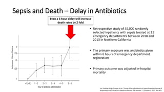
  • 31. Sepsis and Death – Delay in Antibiotics • Retrospective study of 35,000 randomly selected inpatients with sepsis treated at 21 emergency departments between 2010 and 2013 in Northern California • The primary exposure was antibiotics given within 6 hours of emergency department registration • Primary outcome was adjusted in-hospital mortality Even a 6 hour delay will increase death rates by 2 fold Liu, Fielding-Singh, Greene, et al.: Timing of Early Antibiotics in Sepsis American Journal of Respiratory and Critical Care MedicineVolume 196 Number 7 | October 1 2017, 856-863
  • 32. Septic Shock – Fluid Resuscitation • Rescue: During the initial minutes to hours, fluid boluses (a 1- to 2-L fluid bolus of crystalloid/balanced crystalloid solution) are required to reverse hypoperfusion and shock • Optimization: During the second phase, the benefits of giving additional fluid to improve cardiac output and tissue perfusion should be weighed against potential harms • Stabilization: During the third phase, usually 24 to 48 hours after the onset of septic shock, an attempt should be made to achieve a net-neutral or a slightly negative fluid balance • De-escalation: The fourth phase, marked by shock resolution and organ recovery, should trigger aggressive fluid removal strategies Siddharth Dugar et al Sepsis and septic shock: Guideline-based managementCLEVELAND CLINIC JOURNAL OF MEDICINE VOLUME 87 • NUMBER 1 JANUARY 2020
  • 33. Septic Shock – Fluid resuscitation • Within the first 3 hours patients have a lactate level checked, blood cultures drawn prior to antibiotics, broad-spectrum antibiotics given, and 30mL/kg of crystalloid/balanced crystalloid given for hypotension or lactate ≥ 4 mmol/L. • Within 6 hours, vasopressors should be started for hypotension that does not respond to fluid resuscitation to maintain a mean arterial pressure ≥ 65 mm Hg. • Measuring the lactate level is an objective way to assess response to resuscitation, better than other clinical markers, and it continues to be an integral part of sepsis definitions and the Surviving Sepsis Campaign care bundle Jeremy Richards et al. Diagnosis And Management Of Shock In The Emergency Department. Emergency Medicine Practice2014, 16:3 Siddharth Dugar et al Sepsis and septic shock: Guideline-based managementCLEVELAND CLINIC JOURNAL OF MEDICINE VOLUME 87 • NUMBER 1 JANUARY 2020
  • 34. Septic Shock – Source Control • Source control can range from removal of infected intravascular devices to a chest tube for empyema to percutaneous or surgical intervention in cases of cholecystitis and pyelonephritis Siddharth Dugar et al Sepsis and septic shock: Guideline-based managementCLEVELAND CLINIC JOURNAL OF MEDICINE VOLUME 87 • NUMBER 1 JANUARY 2020
  • 35. Septic Shock – Role of corticosteroids • In septic shock, they have a vasopressor-sparing role and reduce the duration of shock, ventilator use, and ICU stay • However, the evidence is not conclusive that giving corticosteroids for sepsis improves clinical outcomes or survival • They can be added as adjunctive therapy for patients requiring higher doses of vasopressors • If corticosteroids are used in septic shock, current guidelines recommend hydrocortisone 200 mg per day intravenously as a continuous drip or 50 mg bolus in 4 divided doses for at least 3 days Siddharth Dugar et al Sepsis and septic shock: Guideline-based managementCLEVELAND CLINIC JOURNAL OF MEDICINE VOLUME 87 • NUMBER 1 JANUARY 2020
  • 36. Septic Shock – Biomarkers • C-reactive protein and erythrocyte sedimentation rate have been used in the past, but with limited success • Procalcitonin has emerged as a method to help detect bacterial infections early and to guide de-escalation or discontinuation of antibiotics. • Galactomannan and beta-D-glucan can be used to detect infections with fungi, specially Aspergillus. Beta-d-glucan is more sensitive for invasive Aspergillus, while galactomannan is more specific Siddharth Dugar et al Sepsis and septic shock: Guideline-based managementCLEVELAND CLINIC JOURNAL OF MEDICINE VOLUME 87 • NUMBER 1 JANUARY 2020
  • 37. Obstructive shock Due to obstruction of great vessels or heart that impedes the blood flow Causes: - cardiac tamponade; tension pneumothorax; pulmonary or air embolism Mangement: - Prompt relief of obstruction: e.g. pericardiocentesis for tamponade, chest drain for tension pneumothorax - Fluid and inotrope are for temporary support Department of Anaesthesia and Intensive Care, the Chinese Universityof Hong Kong July 2015
  • 38. Pitfalls Jeremy Richards et al. Diagnosis And Management Of Shock In The EmergencyDepartment. EmergencyMedicine Practice 2014, 16:3