Common pitfalls in bone marrow
biopsy based diagnostic approach
Dr. N. Varma
Prof. & Head - Hematology
PGIMER, Chandigarh, India
Bone marrow (BM) examination
• Gold standard investigation for diagnosing and
monitoring many hematological diseases
• Useful for investigating various non-hematological
conditions
• Combination of bone marrow aspirate and trephine
biopsy: fine cytological detail, the organization of
BM, and the presence of focal abnormalities
Good-to-have Information
• Accurate clinical information; context and questions being
asked; details of previous investigations
• For neoplastic diseases: ? primary diagnostic investigation/
staging procedure/ re-examination to assess response to
treatment (including transplantation)
• The type and timing of previous BM transplantation are
also important factors; kinetics of engraftment differ
between conditioning regimes and graft types
• Knowledge of the recent therapeutic use of growth factors
such as G-CSF; these may transiently have major modifying
effects on hemopoiesis that can mask or mimic genuine
pathology
Pitfalls in obtaining and interpreting
bone marrow aspirates
• BM aspiration done when not needed
• BM aspiration not done when needed
• BM aspiration done on the wrong site
• The clinical context not adequately assessed and the
correct range of tests is therefore not done on the aspirate
• False negative result as a consequence of a sampling error
• The aspirate is not interpreted together with the trephine
biopsy sections
• The aspirate is misinterpreted
– Problems relating to technical quality
– Correct stains not performed
– Features present not noted
– Misinterpretation of an adequate aspirate
Limiting factors for interpretation of BMB
• Inadequate clinical, hematological (blood and aspirate
findings), genetic and radiological information
• Inadequate specimen
– Too small
– Too crushed/distorted
– Both
– Poorly decalcified/processed
• Inadequate sections (thickness, number of levels)
• Inadequate stains (poor technical quality, range too limited)
• Insufficient experience to avoid common pitfalls
(eg, differential diagnosis of granulomas or fibrosis)
• Insufficient confidence to avoid concluding ‘consistent with’
• ‘Invisible’ pathology
• Forgetting to look at the bone trabeculae and stroma
Common Ancillary Studies complementary to
Bone Marrow Morphologic Examination
• Cytogenetics on BM aspirate or peripheral blood
sample
• FISH studies on BM aspirate or touch preparations
• Molecular studies (PCR or RT-PCR) to detect specific
translocations and/or antigen receptor gene
rearrangements
• Flow cytometric Immunophenotyping of BM aspirate
or peripheral blood cells
• Immunohistochemistry on paraffin sections
• Enzyme cytochemistry on marrow aspirate or
peripheral smear slides
A systematic approach to diagnosis is
required for:
• D/D of hypoplasia/aplasia
• D/D of megaloblastic hemopoiesis
• Assessing key histological features of myelodysplastic
and myeloproliferative haemopoiesis
• D/D of bone marrow fibrosis
• Assessing patterns of lymphoid infiltration associated
with various lymphomas, especially small B-cell
lymphomas
• D/D of granulomatous pathologies
1. D/D hypocellular marrow
Normocellular
• CBC and reticulocyte count
• Blood film examination
• Bone marrow aspirate and trephine biopsy
• HbF% in children
• Peripheral blood lymphocyte cultures for clastogens induced
chromosomal breakage studies
• Ham’s test and / or flowcytometry for GPI anchored proteins
• Urine hemosiderin (if Ham’s test and / or FCM for GPI anchored +)
• Vitamin B12 and folate levels
• Liver function tests
• Renal function tests
• Viral markers (hepatitis A, B, C; EBV; CMV; HIV)
• Antinuclear antibody and anti ds-DNA
• Chest x-ray
• Abdominal ultrasound scan
Investigations recommended for suspected AA
CBS 958
Varma N et al. Multiple constitutional aetiological factors in BMFS patients… Indian J Med Res 2006
Fanconi Anemia associated Aplastic Anemia
(A-1366/11; Tx-1204/11)
2. Subtle increase of immature cells: in hypocellular marrow ?leukemia/lymphoma
IHC: Blasts positive for CD34, anti-MPO (A-1366/11; Tx-1204/11)
Diagnosis: Hypocellular AML
2. Subtle increase of immature cells in hypocellular marrow
3. Problem in differentiating AML-M6 and megaloblastic anemia
Megaloblastic anemia ? Megaloblastic anemia
3. Problem in differentiating AML-M6 and megaloblastic anemia
(A-1305/10; Tx-1056/10) IHC: Blasts positive for anti-MPO, CD34
AML- M6
Hb TLC Platelet
count
Reticulocyte DLC PBF
7 g/dl 7.3 x
109/L
7 x 10⁹/L 5.36% P60L34M4E2 Moderate anisocytosis,
microcytes, macrocytes,
hypochromia & polychromasia
3. D/D of ‘megaloblastic anemia’ picture
Bone marrow aspirate
(BM A-1404/12)
Cellular
M:E= 1:1
Blast1, Pm2, My 40 ,Mm 1, P44, L8, M1, E2
Megakaryocytes: Adequate
Bone marrow
biopsy
(T-1226/12)
Mildly hypercellular with relative erythroid hyperplasia with
megaloblstic changes. Granulocytes and megakaryocytes are adequate.
Common pitfalls in bone marrow biopsy based diagnostic approach
Granulocytes
Granulocytes
Monocytes
RBCs
Final diagnosis: Classical PNH
RCMD-RS
3. D/D of ‘megaloblastic anemia’ picture: characterization of ‘MDS’ like pathology.
45 M, bicytopenia
Bone marrow infiltration in a case of Hepatosplenic lymphoma
(A-408/10; Tx-323/10)
4. Pattern of bone marrow infiltration by NHL
Bone marrow intravascular infiltration in a case of Hepatosplenic lymphoma.
IHC for CD34 and CD3 (A-408/10; Tx-323/10)
CD34 CD3
4. Pattern of bone marrow infiltration by NHL
Plasma cells in a case of Multiple myeloma (A-1441/08; Tx-1161/08)
5. Differentiation between reactive and malignant plasma cells
IHC: Kappa light chain
Reactive plasmacytosis + LD bodies
(A-1535/11; Tx-1356/11)
5. Differentiation between reactive and malignant plasma cells
Reactive increase in plasma cells in a case of Tubercular Granuloma
(A-1198/12; Tx-1042/12)
5. Differentiation between reactive and malignant plasma cells
BM: GranulomaAFB stain
Renal Osteodystrophy
(A-391/12; Tx-339/12)
6. Identification of etiology in fibrosis
Acute panmyelosis with myelofibrosis
(A-185/13; Tx-167/13)
ALL with fibrosis
(A-1476/12; Tx-1288/12)
7. Identification of subtle infiltration of leukemia/lymphoma in fibrotic marrow
IHC for CD34
7. Identification of subtle infiltration of leukemia/lymphoma in fibrotic marrow;
IHC is required
IHC for TdT
IHC for CD20
ALL with fibrosis
(A-1476/12;
Tx-1288/12)
Amyloid deposition in vessel wall
(A-1554/12; Tx-1356/12)
Congo Red stain
8. Problem in cases with subtle Amyloid deposition- need to be confirmed by special
staining by Congo Red
9. Problem in assigning myelodysplasia as reactive or primary
RCMD- predominantly dysplastic megakaryocytes (A-803/12; Tx-690/12)
9. Problem in assigning myelodysplasia as reactive or primary
Case with sepsis- dysplastic megakaryocytes
(A-55/12; Tx-51/12)
Metastatic carcinoma of GIT (A-105/13; Tx-93/13)
10. Problem in assessment of focal lesions- like metastasis may be missed if
sample is inadequate, and also in identification of primary site. These non-
hematologic malignancies may mimic hematological malignancies also.
Metastatic carcinoma- Prostate
(A-983/09; Tx-763/09)
10. Problem in assessment of focal lesions- like metastasis may be missed if
sample is inadequate, and also in identification of primary site. These non-
hematologic malignancies may mimic hematological malignancies also.
Granuloma- TB (A-1198/12; Tx-1042/12)
11. Problem in assessment of focal lesions- like granuloma may be missed if
inadequate sample and also in differentiation of granuloma etiology
Granuloma- Hodgkin’s Lymphoma
(A-1252/12; Tx-1091/12)
11. Problem in assessment of focal lesions- like granuloma may be missed if
inadequate sample and also in differentiation of granuloma etiology
12. Problem in cases with only necrosis- where etiology can not be assessed
BM Necrosis- (A-330/11; Tx-286/11)
13. Problem in identification of lymphocytosis, esp in NK/ T-cell infiltration as
reactive increase or malignant
NK leukemia/lymphoma
(A-444/12; Tx-314/12)
Bone Marrow Trephine Biopsy 314/12
Splenectomy section (S-12985/12) of this case.
IHC for CD56 highlighting NK cell increase in spleen;
case with increased lymphocytes on bone marrow.
13. Problem in identification of lymphocytosis, esp in NK/ T-cell infiltration as
reactive increase or malignant
14. Problem in differentiation of syntitial variant of Hodgkin’s lymphoma and ALCL
Reported as Anaplastic large cell lymphoma
(A-1255/08; Tx-1020/08)
IHC for CD30
IHC for CD15
14. Problem in differentiation of syntitial variant of Hodgkin’s lymphoma and ALCL-
IHC required for differentiation
Reported as Anaplastic large cell lymphoma
(A-1255/08; Tx-1020/08)
15. Problem in identification T-cell rich B-cell lymphoma
IHC for CD3 IHC for CD20
16. There can be technical artefacts leading to inconclusive findings
Washed off marrow spaces
Washed off marrow spaces, hemorrhage and cartilage
16. Procedural artefacts leading to inconclusive findings
16. Technical artefacts leading to inconclusive findings
Crushed marrow spaces
17. 15yrs/M,
TLC 32x109/L, Blasts 95%
Scanty aspirate smears
Tx BxPB smear
MPO +
Positive Markers: CD13, CD33, Anti
MPO, CD19, CD10, CD34, CD45, CD123, H
LADR
Negative Markers: T lineage
FCM-IP Diagnosis: Mixed Phenotype Acute Leukemia (B/ Myeloid)
1444 bp
943 bp
754 bp
585 bp
458 bp
341 bp
258 bp
NC P1 P2 P3 P4 P5 P6 M
bcr-abl transcripts in 1 MPAL (P1) and 5 different CML (P2-6) patients
b3a2 – 385 bp
b2a2 – 310 bp
Bhatia P, Binota J, Varma N, Bansal D, Trehan A, Marwaha RK, Malhotra P, Varma S. A Study on the
Expression of BCR-ABL Transcript in Mixed Phenotype Acute Leukemia (MPAL) Cases Using the
Reverse Transcriptase Polymerase Reaction Assay (RT-PCR) and its Correlation with Hematological
Remission Status Post Initial Induction Therapy. Mediterr J Hematol Infect Dis. 2012;4(1):e2012024.
18. 22M, DOA: 19.11.06; DOD: 26.11.06
C/O fever (↑↑-↑↑↑) 4 weeks
PUO: ? Disseminated TB / lymphoreticular malignancy
CBC: Hb 8.7 g/dl, Retics 1.6-2.6%, TLC 1800-700/ l, platelets 27000-12000/ l; PTI 86%
Bilirubin 4.2/2.8, 4.9/3.6; Serum ferritin 7557.8 mg/dl; Fibrinogen 2.85 g/l; TG 410
BM no. A-1083/06
EBV Zebra LMP 1 EBNA
HPS / HLH
•Screen for underlying genetic, autoimmune,
infectious and malignant diseases
•Uncontrolled hypercytokinemia & many triggers
•Early diagnosis and Rx
PM no. 21703
19. 5 ys. FCh; AML with increased mast cells/ basophils
MPO cytochemistry
P 30 / 07: CD 117 (APAAP)
Varma N, Varma S, Wilkins B.
Br J Haematol 2000;111:991.
Mast cell tryptase
AML with mastocytosis [Systemic mastocytosis with associated
clonal hematological non-mast cell disease (SM-AHNMD)]
Few representative examples
• Assessment of focal lesions
• Differentiation between reactive lymphoid infiltrate and NHL
• Differentiation between reactive and malignant plasma cells
• Identification of malignancies with associated fibrosis
• Effect of growth factors
• Differentiation between hematogones and blasts
• Differentiation between megaloblastic anemia and acute leukemia
• Differentiation between aplastic bone marrow and hypoplastic
myelodysplastic syndrome or hypoplastic acute leukemia
• Identification of lymphomas having a tendency for intravascular
infiltration in the BM
• Subtle amyloid deposition
• Differentiation of macrophage infiltrates and other pathologies that
resemble granulomatous infiltration
• Procedure related artefacts
Take home message
• Integration of clinical, laboratory and imaging information
• Not to assess histology in isolation; uni- / bilateral bx; dry aspirate
• Components of an integrated approach to interpretation are:
– Adequate size of trephine core, with minimal disruption by trauma caused
during collection.
– Access to detailed clinical information and results of additional tests
(specially, peripheral blood cell counts, blood and BM aspirate
cytomorphology, flow cytometry, cytogenetic analysis and radiological
imaging).
– Systematic assessment of all BM components, including trabecular bone and
interstitial stroma.
– Awareness of pathologies that may be ‘invisible’ in trephine specimens
without immunostaining.
– Use of preselected antibody panels for immunostaining and familiarity with
the expected results, including controls.
– Experience in interpreting additional molecular studies, such as clonality PCR
and fluorescence in-situ hybridization.
– Familiarity with the major patterns of bone marrow involvement by reactive
and neoplastic conditions and their differential diagnosis.
– A collaborative approach to working with diverse clinical and laboratory
colleagues.
– Ideally, hematopathologists should report BM Bx along with BM aspirate.
Thank You
Common pifalls in BMB interpretation can be avoided

More Related Content

PPTX
Giant cell lesion.pptx
PPTX
ROLE OF FLOW CYTOMETRY IN LEUKEMIAS
PPTX
CNS papillary neoplasm
PPTX
Dr shashi bansal approch to bone marrow examination
PPTX
Myelodysplastic Syndrome.pptx
PPTX
Minimal Residual Disease in leukaemia andhematological malignancies
PPTX
Acute myeloid leukemia
PPTX
Muscle biopsy interpretation
Giant cell lesion.pptx
ROLE OF FLOW CYTOMETRY IN LEUKEMIAS
CNS papillary neoplasm
Dr shashi bansal approch to bone marrow examination
Myelodysplastic Syndrome.pptx
Minimal Residual Disease in leukaemia andhematological malignancies
Acute myeloid leukemia
Muscle biopsy interpretation

What's hot (20)

PPTX
Pitfalls in diagnosis of soft tissue tumors of childhood
PPTX
papillary lesions of the breast.pptx
PPTX
Endothelium in health and diseases
PDF
The 5th edition of the World Health Organization Classification of Haematolym...
PPTX
Pseudosarcoma
PPTX
Approach to lymphoma
PPTX
Renal pediatric tumors
PPTX
Giant cell lesions of bone
PDF
Mature T/NK cell Neoplasms
PPTX
approach to lymph node cytology part 2
PPTX
Minimal residual disease in AML
PPTX
The Paris System for Reporting Urinary Cytology
PPTX
Small round cell tumors
PPTX
Myelodysplastic Syndrome
PPTX
Minimal Residual Disease in Acute lymphoblastic leukemia
PPTX
Recent changes in 2016 who classification of myeloid neoplasm
PPTX
Microsatellite instability
PPTX
IMWG updates on plasma cell dyscrasias
PPTX
Pitfalls in soft tissue tumor diagnosis
PPTX
Epithelial and mesenchymal transition in invasion and metastasis
Pitfalls in diagnosis of soft tissue tumors of childhood
papillary lesions of the breast.pptx
Endothelium in health and diseases
The 5th edition of the World Health Organization Classification of Haematolym...
Pseudosarcoma
Approach to lymphoma
Renal pediatric tumors
Giant cell lesions of bone
Mature T/NK cell Neoplasms
approach to lymph node cytology part 2
Minimal residual disease in AML
The Paris System for Reporting Urinary Cytology
Small round cell tumors
Myelodysplastic Syndrome
Minimal Residual Disease in Acute lymphoblastic leukemia
Recent changes in 2016 who classification of myeloid neoplasm
Microsatellite instability
IMWG updates on plasma cell dyscrasias
Pitfalls in soft tissue tumor diagnosis
Epithelial and mesenchymal transition in invasion and metastasis
Ad

Viewers also liked (20)

PPTX
Bone marrow biopsy and interpretation
PPTX
Bone marrow aspiration
PPT
Bone marrow aspiration and biopsy
PPT
Bone marrow morphology
PPTX
Common pitfalls in bone marrow biopsy based diagnostic approach
PPT
Bone marrow class
PPTX
processing of bone marrow trephine biopsy
PPTX
Bone marrow aspiration
PPTX
Bone marrow aspirate&biopsy preparation
PPT
Bm examination
PPTX
Acute leukemia
PPT
Bone marrow
PDF
Pavuls presentation bonemarrow aspiration techniques
PPT
Acute Pancreatitis
PPTX
Haemopoisis and Histology of Bone Marrow
PPTX
Anemia aplasica adquirida
PDF
Alterações
PPT
Anemia
PPT
3..rafi ghori megaloblastic anaemia
PPT
Dlc by peripheral smear comparison with automated differential
Bone marrow biopsy and interpretation
Bone marrow aspiration
Bone marrow aspiration and biopsy
Bone marrow morphology
Common pitfalls in bone marrow biopsy based diagnostic approach
Bone marrow class
processing of bone marrow trephine biopsy
Bone marrow aspiration
Bone marrow aspirate&biopsy preparation
Bm examination
Acute leukemia
Bone marrow
Pavuls presentation bonemarrow aspiration techniques
Acute Pancreatitis
Haemopoisis and Histology of Bone Marrow
Anemia aplasica adquirida
Alterações
Anemia
3..rafi ghori megaloblastic anaemia
Dlc by peripheral smear comparison with automated differential
Ad

Similar to Common pitfalls in bone marrow biopsy based diagnostic approach (20)

PPTX
bone marrow.pptx
PPTX
ACUTE LEUKEMIA CME FINAL............pptx
PPTX
Hematological Malignancies 1/2
PDF
peripheral smear anemea clues.pdf
PPT
Cases 2
PPTX
Approach to thrombocytopenia.pptx
PPTX
Investigations of pancytopenia
PPT
Pancytopenia Approach
PPTX
Pancytopenia
PPTX
1_Dr Jasmita Das Peripheral blood smear examination with Romanowsky stains.pptx
PPT
WHO 2008 classification of hematolymphoid neoplasms.ppt
PPTX
Flowcytometry
PPTX
Ophthalmic manifestations of leukemia
PDF
Myelodysplastic Syndromes (MDS)
PPTX
Hematological Malignancies-leukemias.pptx
PDF
Leukemia and lymphoma PATHOLOGY REVISION NOTES
PDF
Hemoblastosis lecture by pathological anatomy
PPTX
Lymphoma spillover.pptx
PPTX
Myelodysplastic syndrome
PPT
A Case of Primary Myelofibrosis
bone marrow.pptx
ACUTE LEUKEMIA CME FINAL............pptx
Hematological Malignancies 1/2
peripheral smear anemea clues.pdf
Cases 2
Approach to thrombocytopenia.pptx
Investigations of pancytopenia
Pancytopenia Approach
Pancytopenia
1_Dr Jasmita Das Peripheral blood smear examination with Romanowsky stains.pptx
WHO 2008 classification of hematolymphoid neoplasms.ppt
Flowcytometry
Ophthalmic manifestations of leukemia
Myelodysplastic Syndromes (MDS)
Hematological Malignancies-leukemias.pptx
Leukemia and lymphoma PATHOLOGY REVISION NOTES
Hemoblastosis lecture by pathological anatomy
Lymphoma spillover.pptx
Myelodysplastic syndrome
A Case of Primary Myelofibrosis

More from spa718 (20)

PPTX
1600 1620 siwanon jirawatnotai
PPTX
Controversies in hepato-biliary surgery
PPTX
Controversies in Colorectal Cancer
PPT
Pancreatic Cancer
PPT
Chemoradiation vs Surgery for rectal cancer
PPTX
Cholangiocarcinoma
PPTX
Immunotherapy for Colorectal Cancer
PPTX
Surgical Approach to Non Small Cell Lung Cancer
PPTX
Role of Radiation Therapy for Lung Cancer
PDF
Update on Management of Triple Negative Breast Cancer
PPTX
Technical Advances in radiotherapy for Lung (and liver) Cancer
PPT
Controversies in Surgical Approach to Breast Cancer
PPTX
ImmunoOncology in Lung Cancer
PDF
Breast Cancer Highlights: ASCO 2015
PPTX
Updates in Radiotherapy for Breast Cancer
PPTX
Regulatory T Cells and GVHD
PPTX
Immunotherapy for Multiple Myeloma
PPT
NHL immunotherapy
PPTX
AML and Cell Therapy
PPTX
Acute Lymphoblastic Lymphoma: Treatment Update
1600 1620 siwanon jirawatnotai
Controversies in hepato-biliary surgery
Controversies in Colorectal Cancer
Pancreatic Cancer
Chemoradiation vs Surgery for rectal cancer
Cholangiocarcinoma
Immunotherapy for Colorectal Cancer
Surgical Approach to Non Small Cell Lung Cancer
Role of Radiation Therapy for Lung Cancer
Update on Management of Triple Negative Breast Cancer
Technical Advances in radiotherapy for Lung (and liver) Cancer
Controversies in Surgical Approach to Breast Cancer
ImmunoOncology in Lung Cancer
Breast Cancer Highlights: ASCO 2015
Updates in Radiotherapy for Breast Cancer
Regulatory T Cells and GVHD
Immunotherapy for Multiple Myeloma
NHL immunotherapy
AML and Cell Therapy
Acute Lymphoblastic Lymphoma: Treatment Update

Recently uploaded (20)

PDF
What if we spent less time fighting change, and more time building what’s rig...
PDF
English Textual Question & Ans (12th Class).pdf
PDF
Paper A Mock Exam 9_ Attempt review.pdf.
PDF
LEARNERS WITH ADDITIONAL NEEDS ProfEd Topic
PDF
LIFE & LIVING TRILOGY - PART - (2) THE PURPOSE OF LIFE.pdf
PDF
CISA (Certified Information Systems Auditor) Domain-Wise Summary.pdf
PPTX
Education and Perspectives of Education.pptx
PPTX
Introduction to pro and eukaryotes and differences.pptx
PDF
LIFE & LIVING TRILOGY- PART (1) WHO ARE WE.pdf
PPTX
Unit 4 Computer Architecture Multicore Processor.pptx
PDF
Τίμαιος είναι φιλοσοφικός διάλογος του Πλάτωνα
PPTX
Computer Architecture Input Output Memory.pptx
PDF
HVAC Specification 2024 according to central public works department
PPTX
Virtual and Augmented Reality in Current Scenario
PDF
Hazard Identification & Risk Assessment .pdf
PPTX
ELIAS-SEZIURE AND EPilepsy semmioan session.pptx
PDF
FOISHS ANNUAL IMPLEMENTATION PLAN 2025.pdf
PDF
MBA _Common_ 2nd year Syllabus _2021-22_.pdf
PPTX
What’s under the hood: Parsing standardized learning content for AI
PDF
LIFE & LIVING TRILOGY - PART (3) REALITY & MYSTERY.pdf
What if we spent less time fighting change, and more time building what’s rig...
English Textual Question & Ans (12th Class).pdf
Paper A Mock Exam 9_ Attempt review.pdf.
LEARNERS WITH ADDITIONAL NEEDS ProfEd Topic
LIFE & LIVING TRILOGY - PART - (2) THE PURPOSE OF LIFE.pdf
CISA (Certified Information Systems Auditor) Domain-Wise Summary.pdf
Education and Perspectives of Education.pptx
Introduction to pro and eukaryotes and differences.pptx
LIFE & LIVING TRILOGY- PART (1) WHO ARE WE.pdf
Unit 4 Computer Architecture Multicore Processor.pptx
Τίμαιος είναι φιλοσοφικός διάλογος του Πλάτωνα
Computer Architecture Input Output Memory.pptx
HVAC Specification 2024 according to central public works department
Virtual and Augmented Reality in Current Scenario
Hazard Identification & Risk Assessment .pdf
ELIAS-SEZIURE AND EPilepsy semmioan session.pptx
FOISHS ANNUAL IMPLEMENTATION PLAN 2025.pdf
MBA _Common_ 2nd year Syllabus _2021-22_.pdf
What’s under the hood: Parsing standardized learning content for AI
LIFE & LIVING TRILOGY - PART (3) REALITY & MYSTERY.pdf

Common pitfalls in bone marrow biopsy based diagnostic approach

  • 1. Common pitfalls in bone marrow biopsy based diagnostic approach Dr. N. Varma Prof. & Head - Hematology PGIMER, Chandigarh, India
  • 2. Bone marrow (BM) examination • Gold standard investigation for diagnosing and monitoring many hematological diseases • Useful for investigating various non-hematological conditions • Combination of bone marrow aspirate and trephine biopsy: fine cytological detail, the organization of BM, and the presence of focal abnormalities
  • 3. Good-to-have Information • Accurate clinical information; context and questions being asked; details of previous investigations • For neoplastic diseases: ? primary diagnostic investigation/ staging procedure/ re-examination to assess response to treatment (including transplantation) • The type and timing of previous BM transplantation are also important factors; kinetics of engraftment differ between conditioning regimes and graft types • Knowledge of the recent therapeutic use of growth factors such as G-CSF; these may transiently have major modifying effects on hemopoiesis that can mask or mimic genuine pathology
  • 4. Pitfalls in obtaining and interpreting bone marrow aspirates • BM aspiration done when not needed • BM aspiration not done when needed • BM aspiration done on the wrong site • The clinical context not adequately assessed and the correct range of tests is therefore not done on the aspirate • False negative result as a consequence of a sampling error • The aspirate is not interpreted together with the trephine biopsy sections • The aspirate is misinterpreted – Problems relating to technical quality – Correct stains not performed – Features present not noted – Misinterpretation of an adequate aspirate
  • 5. Limiting factors for interpretation of BMB • Inadequate clinical, hematological (blood and aspirate findings), genetic and radiological information • Inadequate specimen – Too small – Too crushed/distorted – Both – Poorly decalcified/processed • Inadequate sections (thickness, number of levels) • Inadequate stains (poor technical quality, range too limited) • Insufficient experience to avoid common pitfalls (eg, differential diagnosis of granulomas or fibrosis) • Insufficient confidence to avoid concluding ‘consistent with’ • ‘Invisible’ pathology • Forgetting to look at the bone trabeculae and stroma
  • 6. Common Ancillary Studies complementary to Bone Marrow Morphologic Examination • Cytogenetics on BM aspirate or peripheral blood sample • FISH studies on BM aspirate or touch preparations • Molecular studies (PCR or RT-PCR) to detect specific translocations and/or antigen receptor gene rearrangements • Flow cytometric Immunophenotyping of BM aspirate or peripheral blood cells • Immunohistochemistry on paraffin sections • Enzyme cytochemistry on marrow aspirate or peripheral smear slides
  • 7. A systematic approach to diagnosis is required for: • D/D of hypoplasia/aplasia • D/D of megaloblastic hemopoiesis • Assessing key histological features of myelodysplastic and myeloproliferative haemopoiesis • D/D of bone marrow fibrosis • Assessing patterns of lymphoid infiltration associated with various lymphomas, especially small B-cell lymphomas • D/D of granulomatous pathologies
  • 8. 1. D/D hypocellular marrow Normocellular
  • 9. • CBC and reticulocyte count • Blood film examination • Bone marrow aspirate and trephine biopsy • HbF% in children • Peripheral blood lymphocyte cultures for clastogens induced chromosomal breakage studies • Ham’s test and / or flowcytometry for GPI anchored proteins • Urine hemosiderin (if Ham’s test and / or FCM for GPI anchored +) • Vitamin B12 and folate levels • Liver function tests • Renal function tests • Viral markers (hepatitis A, B, C; EBV; CMV; HIV) • Antinuclear antibody and anti ds-DNA • Chest x-ray • Abdominal ultrasound scan Investigations recommended for suspected AA
  • 10. CBS 958 Varma N et al. Multiple constitutional aetiological factors in BMFS patients… Indian J Med Res 2006 Fanconi Anemia associated Aplastic Anemia
  • 11. (A-1366/11; Tx-1204/11) 2. Subtle increase of immature cells: in hypocellular marrow ?leukemia/lymphoma
  • 12. IHC: Blasts positive for CD34, anti-MPO (A-1366/11; Tx-1204/11) Diagnosis: Hypocellular AML 2. Subtle increase of immature cells in hypocellular marrow
  • 13. 3. Problem in differentiating AML-M6 and megaloblastic anemia Megaloblastic anemia ? Megaloblastic anemia
  • 14. 3. Problem in differentiating AML-M6 and megaloblastic anemia (A-1305/10; Tx-1056/10) IHC: Blasts positive for anti-MPO, CD34 AML- M6
  • 15. Hb TLC Platelet count Reticulocyte DLC PBF 7 g/dl 7.3 x 10⁹/L 7 x 10⁹/L 5.36% P60L34M4E2 Moderate anisocytosis, microcytes, macrocytes, hypochromia & polychromasia 3. D/D of ‘megaloblastic anemia’ picture
  • 16. Bone marrow aspirate (BM A-1404/12) Cellular M:E= 1:1 Blast1, Pm2, My 40 ,Mm 1, P44, L8, M1, E2 Megakaryocytes: Adequate
  • 17. Bone marrow biopsy (T-1226/12) Mildly hypercellular with relative erythroid hyperplasia with megaloblstic changes. Granulocytes and megakaryocytes are adequate.
  • 23. RCMD-RS 3. D/D of ‘megaloblastic anemia’ picture: characterization of ‘MDS’ like pathology. 45 M, bicytopenia
  • 24. Bone marrow infiltration in a case of Hepatosplenic lymphoma (A-408/10; Tx-323/10) 4. Pattern of bone marrow infiltration by NHL
  • 25. Bone marrow intravascular infiltration in a case of Hepatosplenic lymphoma. IHC for CD34 and CD3 (A-408/10; Tx-323/10) CD34 CD3 4. Pattern of bone marrow infiltration by NHL
  • 26. Plasma cells in a case of Multiple myeloma (A-1441/08; Tx-1161/08) 5. Differentiation between reactive and malignant plasma cells IHC: Kappa light chain
  • 27. Reactive plasmacytosis + LD bodies (A-1535/11; Tx-1356/11) 5. Differentiation between reactive and malignant plasma cells
  • 28. Reactive increase in plasma cells in a case of Tubercular Granuloma (A-1198/12; Tx-1042/12) 5. Differentiation between reactive and malignant plasma cells
  • 30. Renal Osteodystrophy (A-391/12; Tx-339/12) 6. Identification of etiology in fibrosis
  • 31. 6. Identification of etiology in fibrosis Acute panmyelosis with myelofibrosis (A-185/13; Tx-167/13)
  • 32. ALL with fibrosis (A-1476/12; Tx-1288/12) 7. Identification of subtle infiltration of leukemia/lymphoma in fibrotic marrow
  • 33. IHC for CD34 7. Identification of subtle infiltration of leukemia/lymphoma in fibrotic marrow; IHC is required IHC for TdT IHC for CD20 ALL with fibrosis (A-1476/12; Tx-1288/12)
  • 34. Amyloid deposition in vessel wall (A-1554/12; Tx-1356/12) Congo Red stain 8. Problem in cases with subtle Amyloid deposition- need to be confirmed by special staining by Congo Red
  • 35. 9. Problem in assigning myelodysplasia as reactive or primary RCMD- predominantly dysplastic megakaryocytes (A-803/12; Tx-690/12)
  • 36. 9. Problem in assigning myelodysplasia as reactive or primary Case with sepsis- dysplastic megakaryocytes (A-55/12; Tx-51/12)
  • 37. Metastatic carcinoma of GIT (A-105/13; Tx-93/13) 10. Problem in assessment of focal lesions- like metastasis may be missed if sample is inadequate, and also in identification of primary site. These non- hematologic malignancies may mimic hematological malignancies also.
  • 38. Metastatic carcinoma- Prostate (A-983/09; Tx-763/09) 10. Problem in assessment of focal lesions- like metastasis may be missed if sample is inadequate, and also in identification of primary site. These non- hematologic malignancies may mimic hematological malignancies also.
  • 39. Granuloma- TB (A-1198/12; Tx-1042/12) 11. Problem in assessment of focal lesions- like granuloma may be missed if inadequate sample and also in differentiation of granuloma etiology
  • 40. Granuloma- Hodgkin’s Lymphoma (A-1252/12; Tx-1091/12) 11. Problem in assessment of focal lesions- like granuloma may be missed if inadequate sample and also in differentiation of granuloma etiology
  • 41. 12. Problem in cases with only necrosis- where etiology can not be assessed BM Necrosis- (A-330/11; Tx-286/11)
  • 42. 13. Problem in identification of lymphocytosis, esp in NK/ T-cell infiltration as reactive increase or malignant NK leukemia/lymphoma (A-444/12; Tx-314/12)
  • 43. Bone Marrow Trephine Biopsy 314/12 Splenectomy section (S-12985/12) of this case. IHC for CD56 highlighting NK cell increase in spleen; case with increased lymphocytes on bone marrow. 13. Problem in identification of lymphocytosis, esp in NK/ T-cell infiltration as reactive increase or malignant
  • 44. 14. Problem in differentiation of syntitial variant of Hodgkin’s lymphoma and ALCL Reported as Anaplastic large cell lymphoma (A-1255/08; Tx-1020/08)
  • 45. IHC for CD30 IHC for CD15 14. Problem in differentiation of syntitial variant of Hodgkin’s lymphoma and ALCL- IHC required for differentiation Reported as Anaplastic large cell lymphoma (A-1255/08; Tx-1020/08)
  • 46. 15. Problem in identification T-cell rich B-cell lymphoma IHC for CD3 IHC for CD20
  • 47. 16. There can be technical artefacts leading to inconclusive findings Washed off marrow spaces
  • 48. Washed off marrow spaces, hemorrhage and cartilage 16. Procedural artefacts leading to inconclusive findings
  • 49. 16. Technical artefacts leading to inconclusive findings Crushed marrow spaces
  • 50. 17. 15yrs/M, TLC 32x109/L, Blasts 95% Scanty aspirate smears Tx BxPB smear MPO +
  • 51. Positive Markers: CD13, CD33, Anti MPO, CD19, CD10, CD34, CD45, CD123, H LADR Negative Markers: T lineage FCM-IP Diagnosis: Mixed Phenotype Acute Leukemia (B/ Myeloid)
  • 52. 1444 bp 943 bp 754 bp 585 bp 458 bp 341 bp 258 bp NC P1 P2 P3 P4 P5 P6 M bcr-abl transcripts in 1 MPAL (P1) and 5 different CML (P2-6) patients b3a2 – 385 bp b2a2 – 310 bp Bhatia P, Binota J, Varma N, Bansal D, Trehan A, Marwaha RK, Malhotra P, Varma S. A Study on the Expression of BCR-ABL Transcript in Mixed Phenotype Acute Leukemia (MPAL) Cases Using the Reverse Transcriptase Polymerase Reaction Assay (RT-PCR) and its Correlation with Hematological Remission Status Post Initial Induction Therapy. Mediterr J Hematol Infect Dis. 2012;4(1):e2012024.
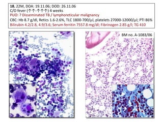
  • 53. 18. 22M, DOA: 19.11.06; DOD: 26.11.06 C/O fever (↑↑-↑↑↑) 4 weeks PUO: ? Disseminated TB / lymphoreticular malignancy CBC: Hb 8.7 g/dl, Retics 1.6-2.6%, TLC 1800-700/ l, platelets 27000-12000/ l; PTI 86% Bilirubin 4.2/2.8, 4.9/3.6; Serum ferritin 7557.8 mg/dl; Fibrinogen 2.85 g/l; TG 410 BM no. A-1083/06
  • 54. EBV Zebra LMP 1 EBNA HPS / HLH •Screen for underlying genetic, autoimmune, infectious and malignant diseases •Uncontrolled hypercytokinemia & many triggers •Early diagnosis and Rx PM no. 21703
  • 55. 19. 5 ys. FCh; AML with increased mast cells/ basophils
  • 57. P 30 / 07: CD 117 (APAAP) Varma N, Varma S, Wilkins B. Br J Haematol 2000;111:991. Mast cell tryptase AML with mastocytosis [Systemic mastocytosis with associated clonal hematological non-mast cell disease (SM-AHNMD)]
  • 58. Few representative examples • Assessment of focal lesions • Differentiation between reactive lymphoid infiltrate and NHL • Differentiation between reactive and malignant plasma cells • Identification of malignancies with associated fibrosis • Effect of growth factors • Differentiation between hematogones and blasts • Differentiation between megaloblastic anemia and acute leukemia • Differentiation between aplastic bone marrow and hypoplastic myelodysplastic syndrome or hypoplastic acute leukemia • Identification of lymphomas having a tendency for intravascular infiltration in the BM • Subtle amyloid deposition • Differentiation of macrophage infiltrates and other pathologies that resemble granulomatous infiltration • Procedure related artefacts
  • 59. Take home message • Integration of clinical, laboratory and imaging information • Not to assess histology in isolation; uni- / bilateral bx; dry aspirate • Components of an integrated approach to interpretation are: – Adequate size of trephine core, with minimal disruption by trauma caused during collection. – Access to detailed clinical information and results of additional tests (specially, peripheral blood cell counts, blood and BM aspirate cytomorphology, flow cytometry, cytogenetic analysis and radiological imaging). – Systematic assessment of all BM components, including trabecular bone and interstitial stroma. – Awareness of pathologies that may be ‘invisible’ in trephine specimens without immunostaining. – Use of preselected antibody panels for immunostaining and familiarity with the expected results, including controls. – Experience in interpreting additional molecular studies, such as clonality PCR and fluorescence in-situ hybridization. – Familiarity with the major patterns of bone marrow involvement by reactive and neoplastic conditions and their differential diagnosis. – A collaborative approach to working with diverse clinical and laboratory colleagues. – Ideally, hematopathologists should report BM Bx along with BM aspirate.
  • 60. Thank You Common pifalls in BMB interpretation can be avoided

Editor's Notes

  • #61: Contribute to diagnose and treat AMLs better!